Department of Veterinary Diagnostic Imaging, College of Veterinary Medicine, Jeonbuk National University, Iksan, Republic of Korea.
Veterinary Diagnostic Laboratory, Michigan State University, Lansing, Michigan, USA.
J Vet Intern Med. 2024 Sep-Oct;38(5):2675-2680. doi: 10.1111/jvim.17192. Epub 2024 Sep 6.
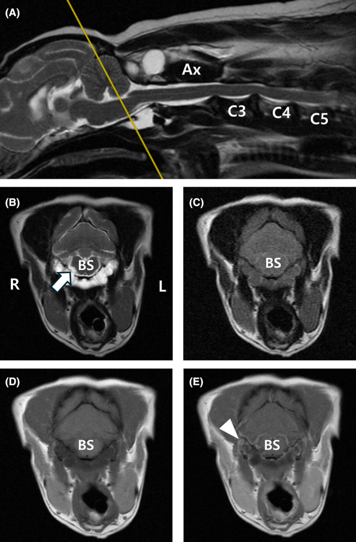
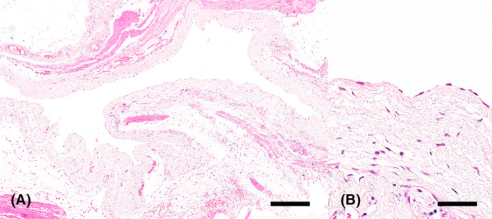
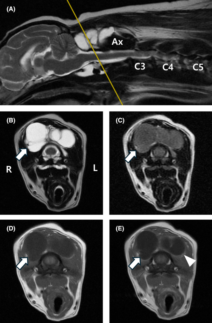
A 14-year-old spayed female Miniature Pinscher presented with tongue curling, dysphagia, hypersalivation, and sublingual gland swelling. Comprehensive evaluation, including neurologic and musculoskeletal examinations, blood work, and urinalysis, revealed no abnormalities other than tongue-related signs. Magnetic resonance imaging (MRI) revealed a multilobed cystic structure in the occipito-atlanto-axial joint, compressing the right hypoglossal canal. The lesion appeared cerebrospinal fluid (CSF)-like on T1-weighted and T2-weighted images, and hyperintense compared with CSF on fluid-attenuated inversion recovery T2-weighted images. The scans suggested mucinous content with enhanced peripheral areas on contrast-enhanced images. Surgical removal and drainage of this cyst were performed, and clinical signs improved markedly. The dorsal cyst was tentatively diagnosed as a ganglion cyst based on histopathologic and imaging findings. Ganglion cysts should be considered in the differential diagnosis for dogs with similar MRI findings and neurologic signs.
一只 14 岁已绝育的雌性迷你宾莎犬出现舌头卷曲、吞咽困难、流涎过多和舌下腺肿胀。除了与舌头相关的症状外,全面评估包括神经和肌肉骨骼检查、血液检查和尿液分析均未发现异常。磁共振成像(MRI)显示寰枕枢关节有多叶囊性结构,压迫右侧舌下神经管。病变在 T1 加权像和 T2 加权像上呈脑脊液样,在液体衰减反转恢复 T2 加权像上与脑脊液相比呈高信号。扫描显示黏液样内容物,增强后呈周边强化。进行了该囊肿的手术切除和引流,临床症状明显改善。根据组织病理学和影像学检查结果,背侧囊肿被初步诊断为神经节囊肿。神经节囊肿应作为具有类似 MRI 表现和神经症状的犬的鉴别诊断之一。