Department of Veterinary Clinical Sciences, College of Veterinary Medicine, The Ohio State University, 601 Vernon Tharp St., Columbus, OH, 43210, USA.
BMC Vet Res. 2019 Nov 6;15(1):396. doi: 10.1186/s12917-019-2152-x.
Extradural intraspinal cysts are fluid accumulations that appear to be associated with increased motion at vertebral joints.
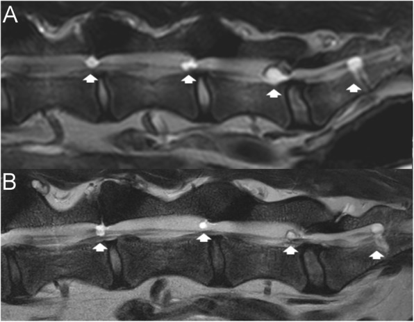
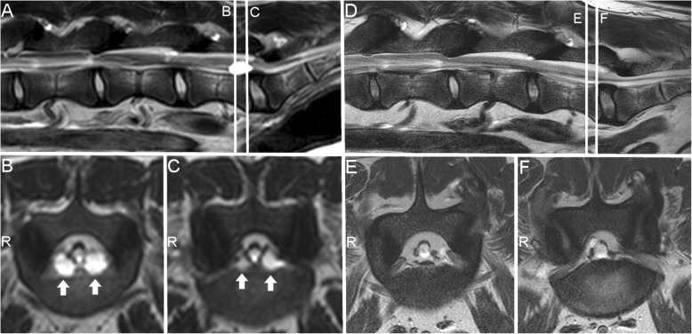
We report the spontaneous regression of lumbar and lumbosacral cysts (presumably synovial cysts) and the unusual occurrence of an S1-2 extradural intraspinal cyst in a dog. The dog presented with lumbosacral pain. Six extradural intraspinal cysts were observed on high-field magnetic resonance imaging from L5-6 to S1-S2. The cysts between L5-6 and L7-S1 ranged from 0.12 to 0.44cm at their largest area. The largest cyst was located at S1-2 (left), measuring 0.84 cm at its largest view. The dog was medically managed. A follow-up magnetic resonance imaging scan was obtained 3.5 years after the first imaging. All cysts except the one at S1-2 had reduced in size. Mean reduction in size was 59.6% (35-81%).
In summary, we report a case with multiple extradural intraspinal cysts that underwent spontaneous regression of all but one cyst during a 3.5-year follow-up period. Whether this is a single occurrence, or is part of the natural history of these cysts in the lumbosacral region of dogs, remains to be established. Spontaneous regression of intraspinal cysts had not been described in dogs.
硬脊膜外脊髓内囊肿是一种液体蓄积,似乎与椎间关节活动增加有关。
我们报告了一例犬腰椎和腰骶部囊肿(推测为滑膜囊肿)的自发消退,以及 S1-2 硬脊膜外脊髓内囊肿的罕见发生。该犬出现腰骶部疼痛。高场磁共振成像显示,从 L5-6 到 S1-S2 共发现 6 个硬脊膜外脊髓内囊肿。L5-6 和 L7-S1 之间的囊肿最大面积为 0.12-0.44cm。最大的囊肿位于 S1-2(左侧),最大径为 0.84cm。该犬接受了药物治疗。首次影像学检查后 3.5 年进行了随访磁共振成像扫描。除 S1-2 处的囊肿外,所有囊肿均缩小。平均缩小率为 59.6%(35%-81%)。
总之,我们报告了一例犬多发性硬脊膜外脊髓内囊肿的病例,在 3.5 年的随访期间,除 S1-2 处的囊肿外,所有囊肿均自发消退。这是单次发生,还是犬腰骶部这些囊肿的自然病史的一部分,尚有待确定。在犬中,脊髓内囊肿的自发消退尚未被描述。