Center for Neurodegeneration and Regeneration, Zilkha Neurogenetic Institute and Department of Physiology and Neuroscience, Keck School of Medicine, University of Southern California, Los Angeles, California 90033
Department of Anesthesiology, Guangzhou Women and Children's Medical Center, Guangzhou Medical University, Guangdong Provincial Clinical Research Center for Child Health, Guangzhou 510623, China.
J Neurosci. 2024 Oct 23;44(43):e0727242024. doi: 10.1523/JNEUROSCI.0727-24.2024.
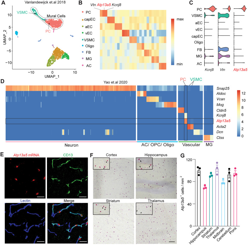
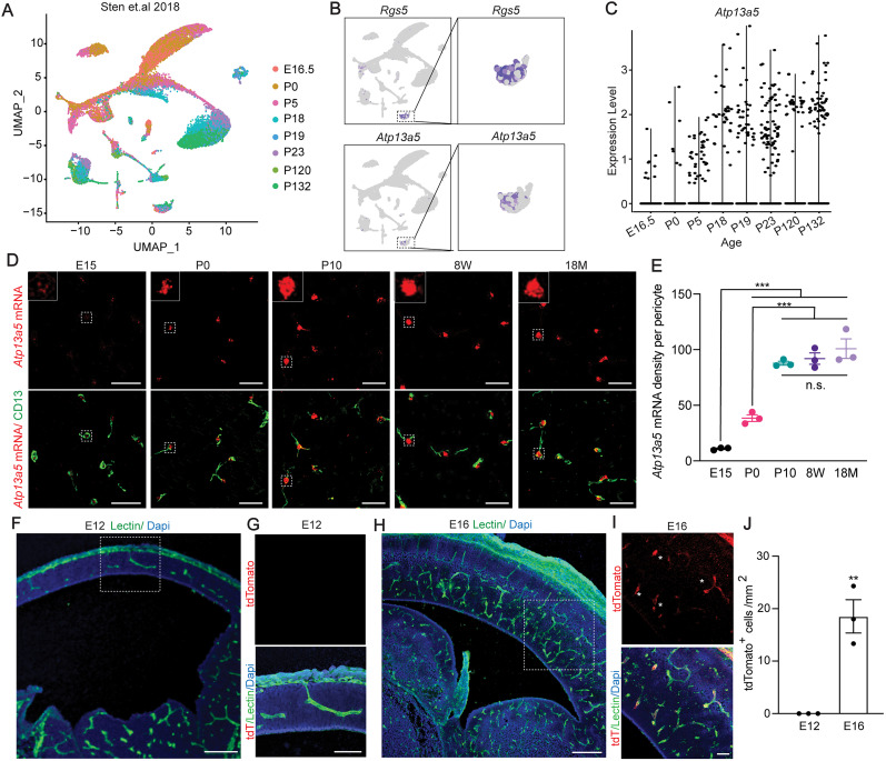
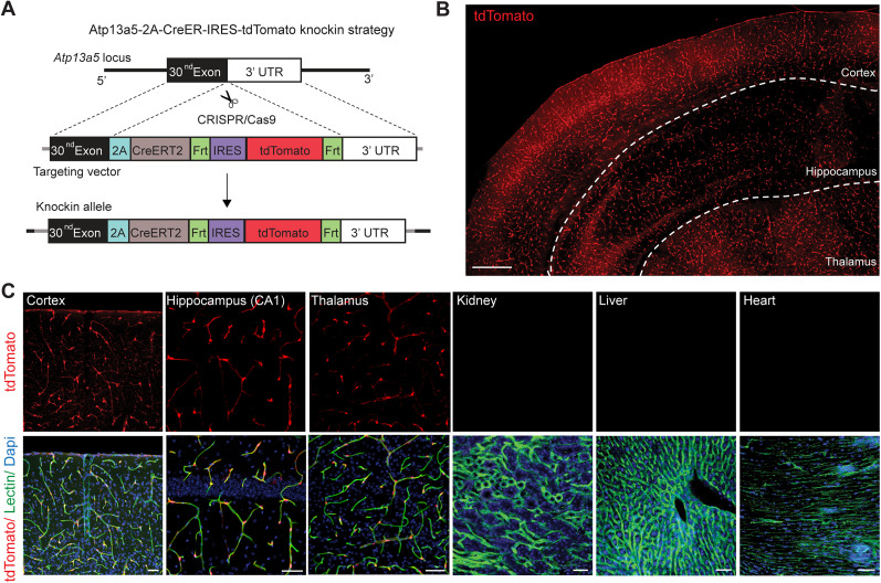
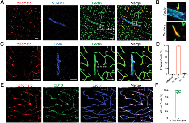
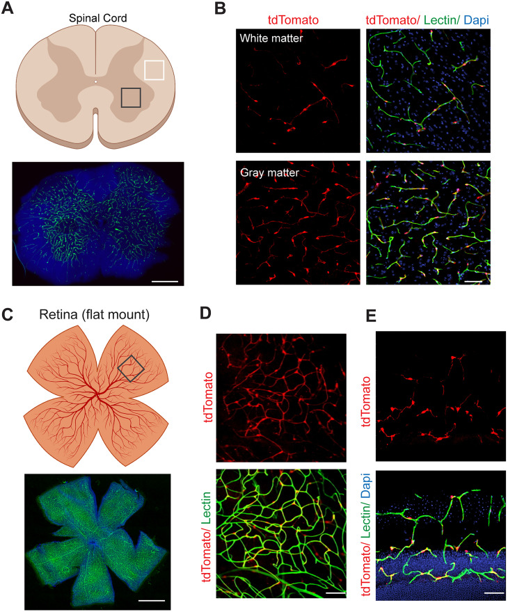
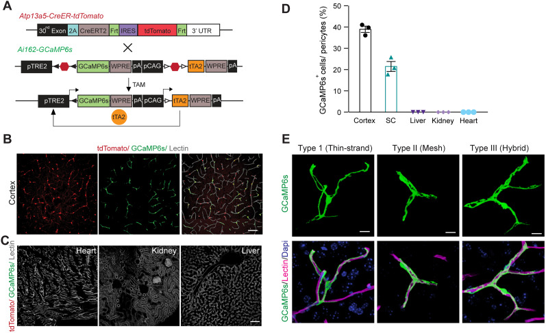
Perivascular mural cells including vascular smooth cells (VSMCs) and pericytes are integral components of the vascular system. In the central nervous system (CNS), pericytes are also indispensable for the blood-brain barrier (BBB), blood-spinal cord barrier, and blood-retinal barrier and play key roles in maintaining cerebrovascular and neuronal functions. However, the functional specifications of pericytes between CNS and peripheral organs have not been resolved at the genetic and molecular levels. Hence, the generation of reliable CNS pericyte-specific models and genetic tools remains very challenging. Here, we report a new CNS pericyte marker in mice. This putative cation-transporting ATPase 13A5 () marker was identified through single-cell transcriptomics, based on its specificity to brain pericytes. We further generated a knock-in model with both tdTomato reporter and Cre recombinase. Using this model to trace the distribution of positive pericytes in mice, we found that the tdTomato reporter reliably labels the CNS pericytes, including the ones in spinal cord and retina but not peripheral organs. Interestingly, brain pericytes are likely shaped by the developing neural environment, as positive pericytes start to appear around murine embryonic day 15 (E15) and expand along the cerebrovasculature. Thus, is a specific marker of CNS pericyte lineage, and this based model is a reliable tool to explore the heterogeneity of pericytes and BBB functions in health and diseases.
血管周细胞包括血管平滑肌细胞(VSMCs)和周细胞,是血管系统的重要组成部分。在中枢神经系统(CNS)中,周细胞对于血脑屏障(BBB)、血脊髓屏障和血视网膜屏障也是不可或缺的,在维持脑血管和神经元功能方面发挥着关键作用。然而,CNS 和外周器官的周细胞在遗传和分子水平上的功能特征尚未得到解决。因此,生成可靠的 CNS 周细胞特异性模型和遗传工具仍然极具挑战性。在这里,我们在小鼠中报告了一种新的 CNS 周细胞标志物。该假定的阳离子转运 ATP 酶 13A5()标志物是通过单细胞转录组学鉴定的,基于其对脑周细胞的特异性。我们进一步生成了一个带有 tdTomato 报告基因和 Cre 重组酶的敲入模型。使用该模型追踪小鼠中阳性周细胞的分布,我们发现 tdTomato 报告基因可靠地标记了 CNS 周细胞,包括脊髓和视网膜中的周细胞,但不包括外周器官中的周细胞。有趣的是,脑周细胞可能受到发育中的神经环境的影响,因为阳性周细胞大约在胚胎第 15 天(E15)开始出现,并沿着脑血管扩张。因此,是 CNS 周细胞谱系的特异性标志物,而这种基于模型的方法是探索健康和疾病中周细胞和 BBB 功能异质性的可靠工具。