Yousufzai S Y, Abdel-Latif A A
Biochem J. 1985 Jun 15;228(3):697-706. doi: 10.1042/bj2280697.
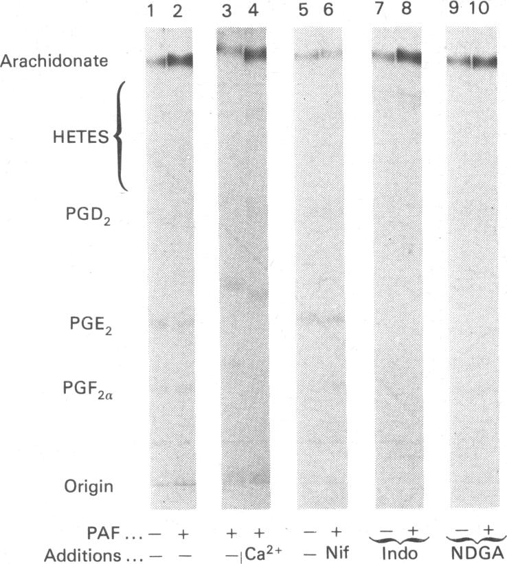
Addition of physiological concentrations (10(-12)-10(-8)M) of platelet-activating factor (PAF) to rabbit iris muscle induced a rapid release (in 15s) of prostaglandin (PG)E2 and 6-oxo-PGF1 alpha, measured by radioimmunoassay and rapid release of 14C-labelled arachidonate and PGE2 in muscle prelabelled with [14C]arachidonic acid, measured by radiochromatography. These PAF actions are concentration- and time-dependent. The effect of PAF on PG release is not mediated through the cyclo-oxygenase pathway. The studies on the properties and mechanism of arachidonate release from phosphatidylinositol and other phospholipids in prelabelled irides by PAF suggest the involvement of a phospholipase A2. This conclusion is supported by the findings: (a) that both the removal of arachidonate and formation of lysophosphatidylinositol, from phosphatidylinositol, by PAF occur concomitantly in a time-dependent manner, (b) that Ca2+ is required for the agonist-induced release of arachidonate and PGE2, and (c) that in contrast to the rapid release of [3H]myo-inositol phosphates by carbachol and other Ca2+-mobilizing agonists previously reported in the iris muscle [Akhtar & Abdel-Latif (1984) Biochem. J. 224, 291-300], PAF (10(-12)-10(-8)M) did not appreciably enhance the release of [14C]myo-inositol phosphates and 32P labelling of phosphatidate and phosphatidylinositol in this tissue. Ca2+-channel antagonists, such as nifedipine, verapamil, diltiazem and manganese inhibited PAF-induced arachidonate and PGE2 release in a dose-dependent manner. K+ depolarization, which causes influx of extracellular Ca2+ in smooth muscle, did not increase the release of arachidonate and PGE2. The ability of Ca2+ antagonists to inhibit arachidonate release by PAF in this tissue probably reflects interference with PAF binding to its receptor. The PAF-induced release of arachidonate and PGE2 occur independently of the cyclo-oxygenase and lipoxygenase pathways. Whether the PAF-induced release of arachidonate and PG in the iris muscle is involved in the pathogenesis of inflammatory and/or physiological reactions in the eye, and how much the inhibitory effects of Ca2+-entry blockers on the PAF actions contribute to the therapeutic use of these drugs, remain to be established.
向兔虹膜肌肉中添加生理浓度(10⁻¹² - 10⁻⁸M)的血小板活化因子(PAF),通过放射免疫测定法可诱导前列腺素(PG)E2和6 - 氧代 - PGF1α迅速释放(15秒内),并且在预先用[¹⁴C]花生四烯酸标记的肌肉中,通过放射色谱法可测得¹⁴C标记的花生四烯酸和PGE2迅速释放。这些PAF的作用具有浓度和时间依赖性。PAF对PG释放的影响不是通过环氧化酶途径介导的。关于PAF从预先标记的虹膜中的磷脂酰肌醇和其他磷脂释放花生四烯酸的特性和机制的研究表明涉及磷脂酶A2。这一结论得到以下发现的支持:(a)PAF从磷脂酰肌醇中去除花生四烯酸并形成溶血磷脂酰肌醇是同时以时间依赖性方式发生的;(b)激动剂诱导的花生四烯酸和PGE2释放需要Ca²⁺;(c)与先前报道的虹膜肌肉中卡巴胆碱和其他Ca²⁺动员激动剂迅速释放[³H]肌醇磷酸不同[阿赫塔尔和阿卜杜勒 - 拉蒂夫(1984年)《生物化学杂志》224,291 - 300],PAF(10⁻¹² - 10⁻⁸M)在此组织中并未明显增强[¹⁴C]肌醇磷酸的释放以及磷脂酸和磷脂酰肌醇的³²P标记。Ca²⁺通道拮抗剂,如硝苯地平、维拉帕米、地尔硫卓和锰,以剂量依赖性方式抑制PAF诱导的花生四烯酸和PGE2释放。K⁺去极化会导致平滑肌中细胞外Ca²⁺内流,但并未增加花生四烯酸和PGE2的释放。Ca²⁺拮抗剂在该组织中抑制PAF诱导的花生四烯酸释放的能力可能反映了其对PAF与其受体结合的干扰。PAF诱导的花生四烯酸和PGE2释放独立于环氧化酶和脂氧合酶途径。PAF在虹膜肌肉中诱导的花生四烯酸和PG释放是否参与眼部炎症和/或生理反应的发病机制,以及Ca²⁺内流阻滞剂对PAF作用的抑制作用在这些药物的治疗应用中有多大贡献,仍有待确定。