Ellsworth Calder R, Chen Zheng, Xiao Mark T, Qian Chaosi, Wang Chenxiao, Khatun Mst Shamima, Liu Shumei, Islamuddin Mohammad, Maness Nicholas J, Halperin Jose A, Blair Robert V, Kolls Jay K, Tomlinson Stephen, Qin Xuebin
Tulane National Primate Research Center, Covington, LA, 70433, USA.
Department of Microbiology and Immunology, Tulane University School of Medicine, New Orleans, LA, 70112, USA.
Cell Mol Life Sci. 2024 Sep 16;81(1):405. doi: 10.1007/s00018-024-05430-w.
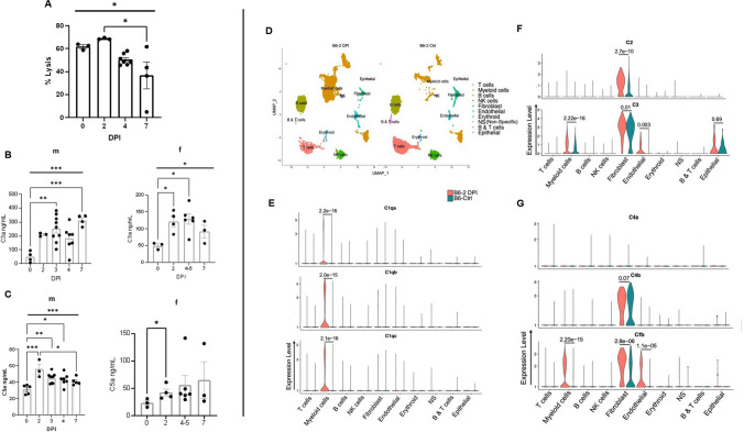
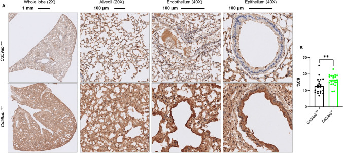
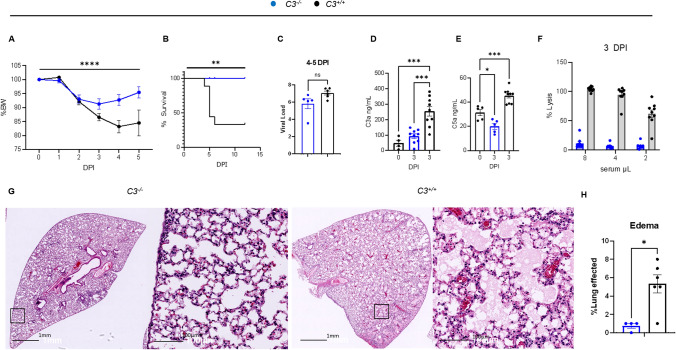
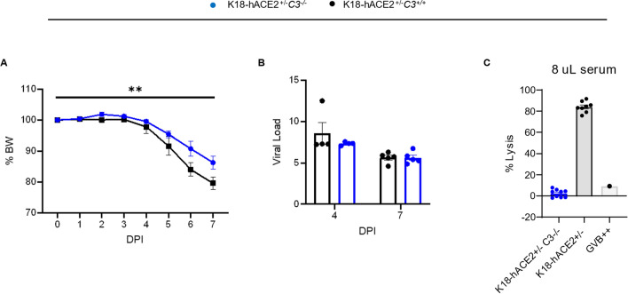
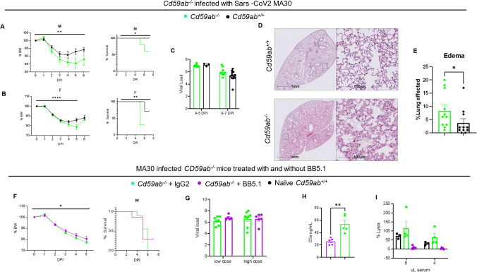
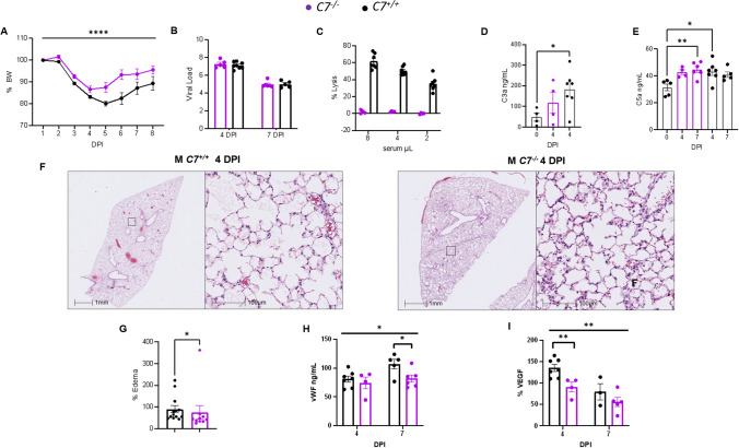
Emerging evidence indicates that activation of complement system leading to the formation of the membrane attack complex (MAC) plays a detrimental role in COVID-19. However, their pathogenic roles have never been experimentally investigated before. We used three knock out mice strains (1. C3; 2. C7; and 3. Cd59ab) to evaluate the role of complement in severe COVID-19 pathogenesis. C3 deficient mice lack a key common component of all three complement activation pathways and are unable to generate C3 and C5 convertases. C7 deficient mice lack a complement protein needed for MAC formation. Cd59ab deficient mice lack an important inhibitor of MAC formation. We also used anti-C5 antibody to block and evaluate the therapeutic potential of inhibiting MAC formation. We demonstrate that inhibition of complement activation (in C3) and MAC formation (in C3. C7, and anti-C5 antibody) attenuates severe COVID-19; whereas enhancement of MAC formation (Cd59ab) accelerates severe COVID-19. The degree of MAC but not C3 deposits in the lungs of C3, C7 mice, and Cd59ab mice as compared to their control mice is associated with the attenuation or acceleration of SARS-CoV-2-induced disease. Further, the lack of terminal complement activation for the formation of MAC in C7 deficient mice protects endothelial dysfunction, which is associated with the attenuation of diseases and pathologic changes. Our results demonstrated the causative effect of MAC in severe COVID-19 and indicate a potential avenue for modulating the complement system and MAC formation in the treatment of severe COVID-19.
新出现的证据表明,补体系统的激活导致膜攻击复合物(MAC)的形成在新冠肺炎中起有害作用。然而,它们的致病作用此前从未经过实验研究。我们使用了三种基因敲除小鼠品系(1. C3;2. C7;3. Cd59ab)来评估补体在重症新冠肺炎发病机制中的作用。C3缺陷小鼠缺乏所有三种补体激活途径的关键共同成分,无法产生C3和C5转化酶。C7缺陷小鼠缺乏MAC形成所需的一种补体蛋白。Cd59ab缺陷小鼠缺乏MAC形成的一种重要抑制剂。我们还使用抗C5抗体来阻断并评估抑制MAC形成的治疗潜力。我们证明,抑制补体激活(在C3中)和MAC形成(在C3、C7和抗C5抗体中)可减轻重症新冠肺炎;而增强MAC形成(Cd59ab)会加速重症新冠肺炎。与对照小鼠相比,C3、C7小鼠和Cd59ab小鼠肺部MAC而非C3的沉积程度与新冠病毒诱导疾病的减轻或加速有关。此外,C7缺陷小鼠中缺乏用于MAC形成的末端补体激活可保护内皮功能障碍,这与疾病和病理变化的减轻有关。我们的结果证明了MAC在重症新冠肺炎中的因果作用,并指出了在重症新冠肺炎治疗中调节补体系统和MAC形成的潜在途径。