F.M. Kirby Research Center for Functional Brain Imaging, Kennedy Krieger Institute, 707 N Broadway, Baltimore, MD, 21205, USA.
Neurosection, Division of MRI Research, Russell H. Morgan Department of Radiology and Radiological Science, Johns Hopkins University School of Medicine, Baltimore, MD, USA.
Fluids Barriers CNS. 2024 Sep 16;21(1):72. doi: 10.1186/s12987-024-00571-3.
Pathways for intravenously administered gadolinium-based-contrast-agents (GBCAs) entering cerebrospinal-fluid (CSF) circulation in the human brain are not well-understood. The blood-CSF-barrier (BCSFB) in choroid-plexus (CP) has long been hypothesized to be a main entry-point for intravenous-GBCAs into CSF. Most existing studies on this topic were performed in animals and human patients with various diseases. Results in healthy human subjects are limited. Besides, most studies were performed using MRI methods with limited temporal resolution and significant partial-volume effects from blood and CSF.
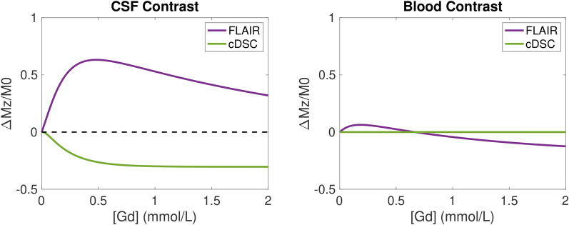
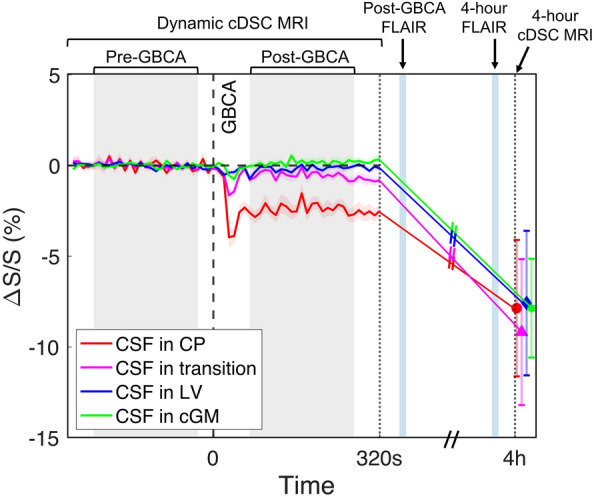
This study employs the recently developed dynamic-susceptibility-contrast-in-the-CSF (cDSC) MRI approach to measure GBCA-distribution in the CSF immediately and 4 h after intravenous-GBCA administration in healthy subjects. With a temporal resolution of 10 s, cDSC MRI can track GBCA-induced CSF signal changes during the bolus phase, which has not been investigated previously. It employs a long echo-time (TE = 1347 ms) to suppress tissue and blood signals so that pure CSF signal is detected with minimal partial-volume effects. GBCA concentration in the CSF can be estimated from cDSC MRI. In this study, cDSC and FLAIR MRI were performed immediately and 4 h after intravenous GBCA administration in 25 healthy volunteers (age 48.9 ± 19.5 years; 14 females). Paired t-tests were used to compare pre-GBCA and post-GBCA signal changes, and their correlations with age were evaluated using Pearson-correlation-coefficients.
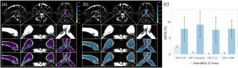
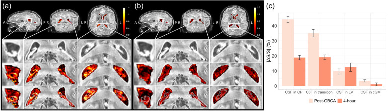
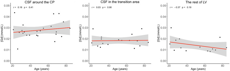
At ~ 20 s post-GBCA, GBCA-induced cDSC signal changes were detected in the CSF around CP (ΔS/S = - 2.40 ± 0.30%; P < .001) but not in the rest of lateral ventricle (LV). At 4 h, significant GBCA-induced cDSC signal changes were observed in the entire LV (ΔS/S = - 7.58 ± 3.90%; P = .002). FLAIR MRI showed a similar trend. GBCA-induced CSF signal changes did not correlate with age.
These results provided direct imaging evidence that GBCAs can pass the BCSFB in the CP and enter ventricular CSF immediately after intravenous administration in healthy human brains. Besides, our results in healthy subjects established a basis for clinical studies in brain diseases exploiting GBCA-enhanced MRI to detect BCSFB dysfunction.
在人类大脑中,静脉注射用钆基对比剂(GBCA)进入脑脊液(CSF)循环的途径尚不清楚。脉络丛(CP)中的血脑屏障(BCSFB)一直被假设为静脉内 GBCA 进入 CSF 的主要入口。该主题的大多数现有研究都是在动物和患有各种疾病的人类患者中进行的。在健康的人类受试者中,结果是有限的。此外,大多数研究都是使用 MRI 方法进行的,这些方法的时间分辨率有限,并且存在来自血液和 CSF 的显著部分容积效应。
本研究采用最近开发的 CSF 中的动态对比敏感度(cDSC)MRI 方法,在健康受试者中测量静脉内 GBCA 给药后立即和 4 小时后 CSF 中的 GBCA 分布。cDSC MRI 的时间分辨率为 10 秒,可在团注阶段跟踪 GBCA 诱导的 CSF 信号变化,这在以前的研究中尚未进行过研究。它采用长回波时间(TE = 1347 ms)来抑制组织和血液信号,从而以最小的部分容积效应检测到纯 CSF 信号。可以从 cDSC MRI 估计 CSF 中的 GBCA 浓度。在这项研究中,在 25 名健康志愿者(年龄 48.9 ± 19.5 岁;14 名女性)中,在静脉内 GBCA 给药后立即和 4 小时进行了 cDSC 和 FLAIR MRI。使用配对 t 检验比较 GBCA 给药前后的信号变化,并使用 Pearson 相关系数评估其与年龄的相关性。
在 GBCA 给药后约 20 秒,在 CP 周围的 CSF 中检测到 GBCA 诱导的 cDSC 信号变化(ΔS/S = -2.40 ± 0.30%;P <.001),但在外侧脑室(LV)的其余部分未检测到。在 4 小时时,在整个 LV 中观察到明显的 GBCA 诱导的 cDSC 信号变化(ΔS/S = -7.58 ± 3.90%;P =.002)。FLAIR MRI 显示出类似的趋势。GBCA 诱导的 CSF 信号变化与年龄无关。
这些结果提供了直接的影像学证据,表明 GBCA 可以穿过 CP 中的 BCSFB,并在健康人脑静脉内给药后立即进入脑室 CSF。此外,我们在健康受试者中的结果为利用 GBCA 增强 MRI 检测 BCSFB 功能障碍的脑部疾病的临床研究奠定了基础。