Lucarelli Nicola Maria, Troise Francesca, Antonicelli Valentina, Greco Sara, Morelli Chiara, Maggialetti Nicola
Interdisciplinary Department of Medicine, Section of Radiology and Radiation Oncology, University of Bari "Aldo Moro", Bari 70124, Italy.
Radiol Case Rep. 2024 Aug 31;19(11):5389-5392. doi: 10.1016/j.radcr.2024.08.001. eCollection 2024 Nov.
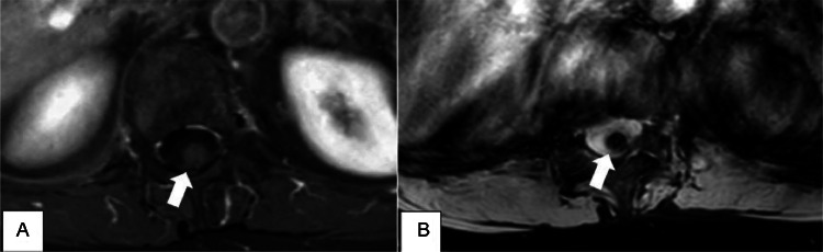
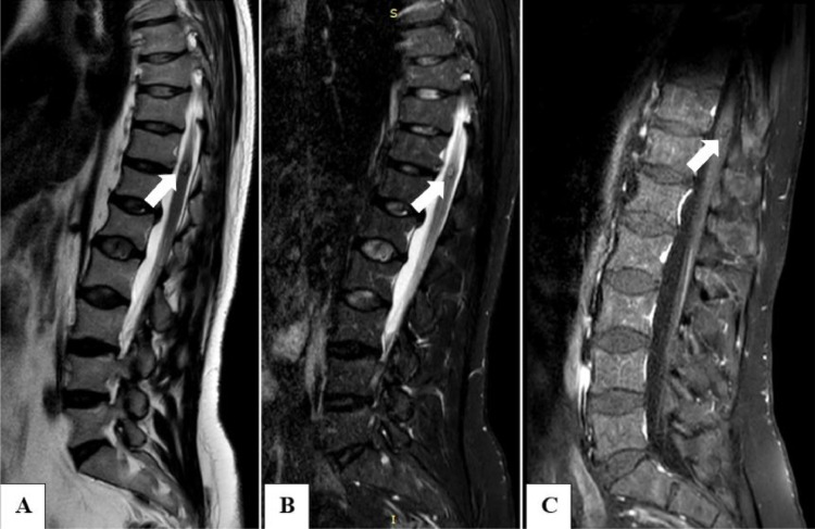
Cavernous malformations are rare vascular anomalies of the central nervous system, occurring in the spinal cord in just 5% of cases. Despite being documented in the literature, intramedullary cavernous malformations are exceedingly rare and often challenging to distinguish from other intramedullary lesions. We report a case of a 42-year-old patient with back pain, right-sided dysesthesias, and impaired proprioception in the distal limbs for approximately 3 months. Magnetic resonance imaging, crucial for differential diagnosis, identified intramedullary cavernous malformations at T11-12. Several conditions can hide the real cause of back pain; however, magnetic resonance imaging can reveal common conditions (such as discal hernia) and rare findings like cavernous malformations. Magnetic resonance imaging remains the study of choice for diagnosing and characterizing intramedullary cavernous malformations.
海绵状血管畸形是中枢神经系统罕见的血管异常,仅5%的病例发生在脊髓。尽管文献中有记载,但髓内海绵状血管畸形极为罕见,且常常难以与其他髓内病变相区分。我们报告一例42岁患者,有背痛、右侧感觉异常以及远端肢体本体感觉受损约3个月。磁共振成像对于鉴别诊断至关重要,其显示在T11 - 12水平存在髓内海绵状血管畸形。有几种情况可能掩盖背痛的真正病因;然而,磁共振成像能够揭示常见情况(如椎间盘突出)以及像海绵状血管畸形这样的罕见发现。磁共振成像仍然是诊断和表征髓内海绵状血管畸形的首选检查方法。