Cardiovascular Translational Research Center, Department of Cell Biology and Anatomy, University of South Carolina, Columbia (SC) 29209, USA.
Department of Pharmacology, Institute of Biomedical Science, University Of Sao Paulo, Sao Paulo (SP) 05508, Brazil.
Function (Oxf). 2024 Nov 20;5(6). doi: 10.1093/function/zqae042.
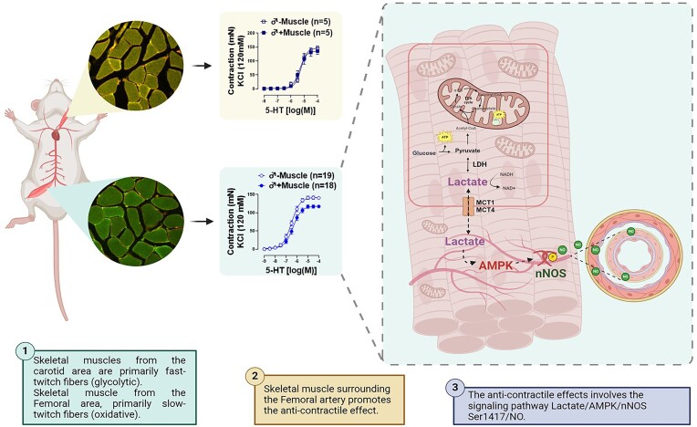
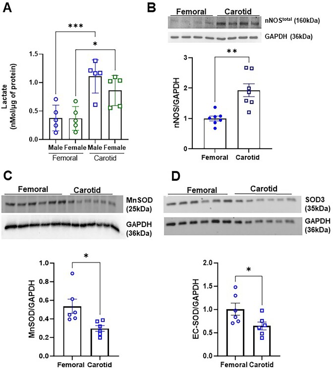
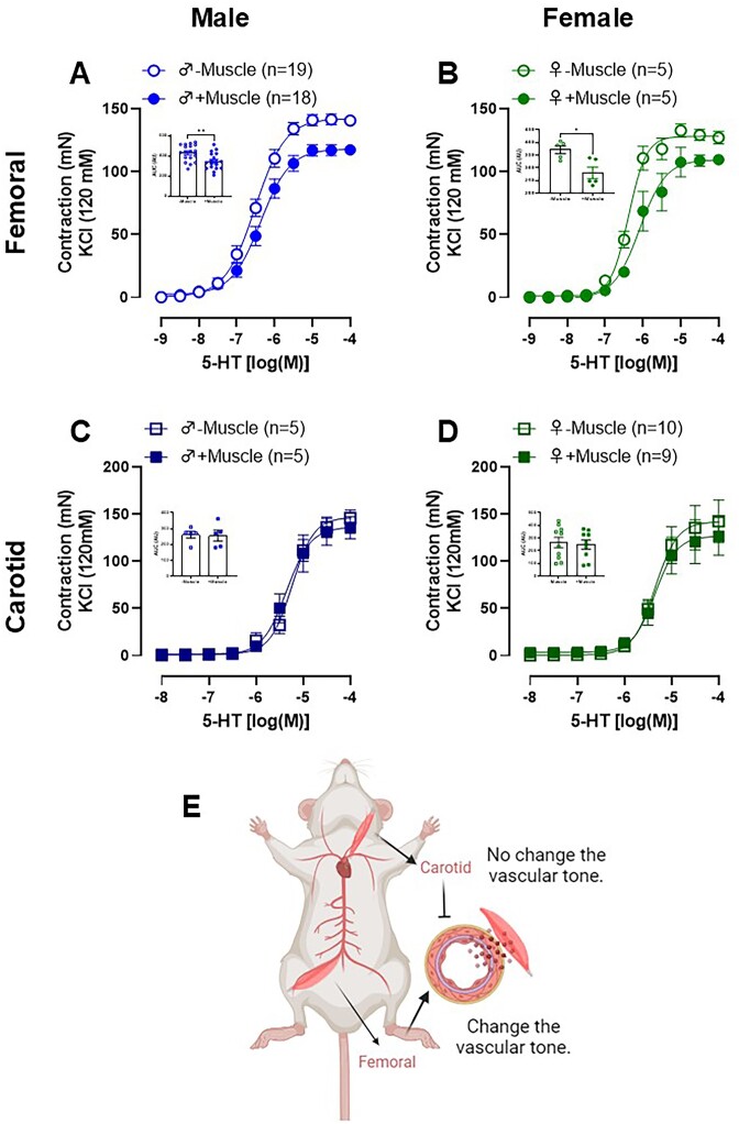
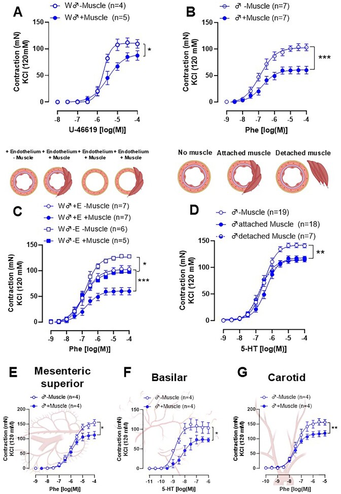
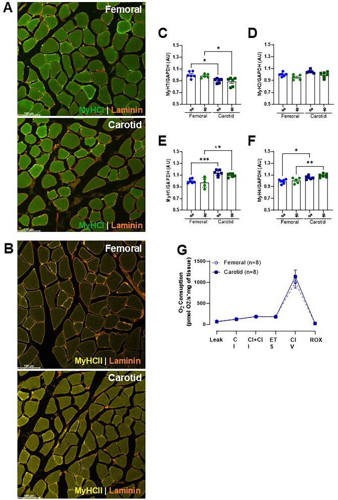
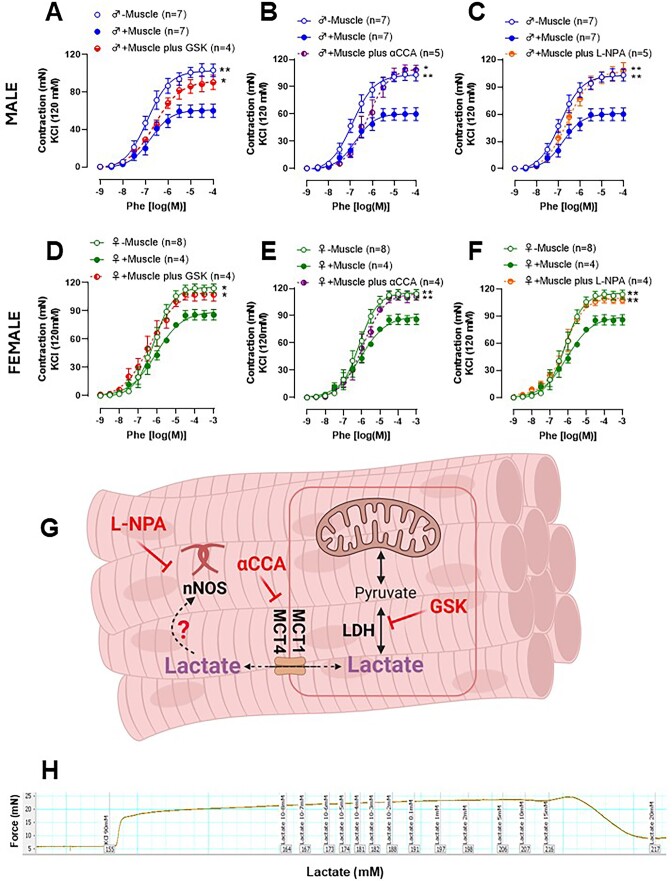
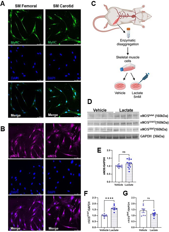
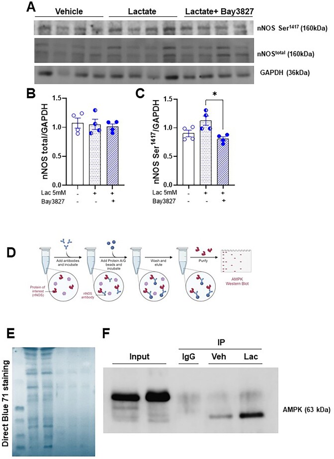
The regulation of vascular tone by perivascular tissues is a complex interplay of various paracrine factors. Here, we investigate the anti-contractile effect of skeletal muscle surrounding the femoral and carotid arteries and its underlying mechanisms. Using male and female Wistar rats, we demonstrated that serotonin, phenylephrine, and U-46619 induced a concentration-dependent vasoconstrictor response in femoral artery rings. Interestingly, this response was diminished in the presence of surrounding femoral skeletal muscle, irrespective of sex. No anti-contractile effect was observed when the carotid artery was exposed to its surrounding skeletal muscle. The observed effect in the femoral artery persisted even in the absence of endothelium and when the muscle was detached from the artery. Furthermore, the skeletal muscle surrounding the femoral artery was able to promote an anti-contractile effect in three other vascular beds (basilar, mesenteric, and carotid arteries). Using inhibitors of lactate dehydrogenase and the 1/4 monocarboxylate transporter, we confirmed the involvement of lactate, as both inhibitors were able to abolish the anti-contractile effect. However, lactate did not directly promote vasodilation; rather, it exerted its effect by activating 5' AMP-activated protein kinase (AMPK) and neuronal nitric oxide synthase (NOS1) in the skeletal muscle. Accordingly, Nω-propyl l-arginine, a specific inhibitor of NOS1, prevented the anti-contractile effect, as well as lactate-induced phosphorylation of NOS1 at the stimulatory serine site (1417) in primary skeletal muscle cells. Phosphorylation of NOS1 was reduced in the presence of Bay-3827, a selective AMPK inhibitor. In conclusion, femoral artery-associated skeletal muscle is a potent paracrine and endocrine organ that influences vascular tone in both sexes. Mechanistically, the anti-contractile effect involves muscle fiber type and/or its anatomical location but not the type of artery or its related vascular endothelium. Finally, the femoral artery anti-contractile effect is mediated by the lactate-AMPK-phospho-NOS1Ser1417-NO signaling axis.
血管周围组织对血管张力的调节是各种旁分泌因子复杂相互作用的结果。在这里,我们研究了股动脉和颈动脉周围骨骼肌的抗收缩作用及其潜在机制。我们使用雄性和雌性 Wistar 大鼠证明,5-羟色胺、苯肾上腺素和 U-46619 在股动脉环中引起浓度依赖性的血管收缩反应。有趣的是,无论性别如何,在存在周围股骨骼肌的情况下,这种反应都会减弱。当颈动脉暴露于其周围的骨骼肌时,没有观察到抗收缩作用。即使在没有内皮细胞的情况下,或者当肌肉与动脉分离时,股动脉中观察到的作用仍然存在。此外,股动脉周围的骨骼肌能够在其他三个血管床(基底动脉、肠系膜动脉和颈动脉)中促进抗收缩作用。使用乳酸脱氢酶和 1/4 单羧酸转运蛋白抑制剂证实了乳酸的参与,因为这两种抑制剂都能够消除抗收缩作用。然而,乳酸本身并没有直接促进血管舒张,而是通过在骨骼肌中激活 5' AMP 激活的蛋白激酶(AMPK)和神经元型一氧化氮合酶(NOS1)来发挥作用。因此,NOS1 的特异性抑制剂 Nω-丙基 L-精氨酸(Nω-Propyl l-arginine),以及在原代骨骼肌细胞中 NOS1 的刺激丝氨酸位点(1417)的磷酸化,阻止了抗收缩作用。在 Bay-3827(一种选择性 AMPK 抑制剂)存在下,NOS1 的磷酸化减少。总之,股动脉相关的骨骼肌是一种强大的旁分泌和内分泌器官,它影响两性的血管张力。从机制上讲,抗收缩作用涉及肌纤维类型和/或其解剖位置,但与动脉类型或其相关的血管内皮无关。最后,股动脉抗收缩作用是由乳酸-AMPK-磷酸化 NOS1Ser1417-NO 信号通路介导的。