University Health Network, Princess Margaret Cancer Centre, Toronto, Ontario, Canada.
University of Toronto, Department of Otolaryngology-Head and Neck Surgery, Toronto, Ontario, Canada.
J Biomed Opt. 2025 Jan;30(Suppl 1):S13706. doi: 10.1117/1.JBO.30.S1.S13706. Epub 2024 Sep 18.
Oral cancer surgery requires accurate margin delineation to balance complete resection with post-operative functionality. Current fluorescence imaging systems provide two-dimensional margin assessment yet fail to quantify tumor depth prior to resection. Harnessing structured light in combination with deep learning (DL) may provide near real-time three-dimensional margin detection.
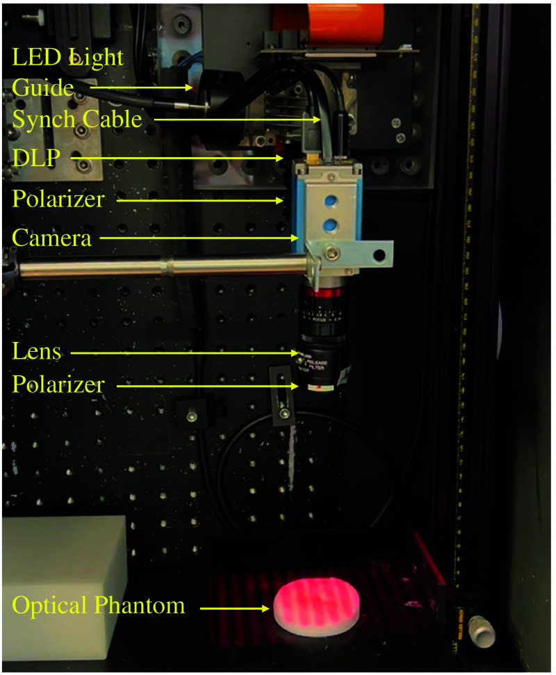
A DL-enabled fluorescence spatial frequency domain imaging (SFDI) system trained with tumor models was developed to quantify the depth of oral tumors.
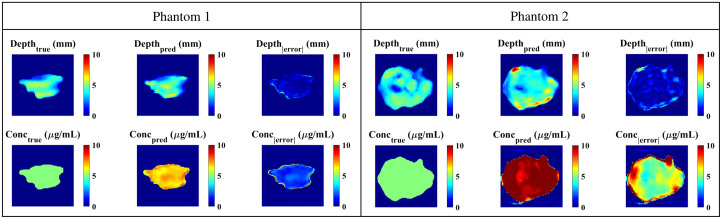
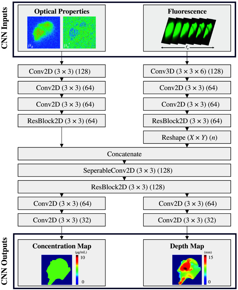
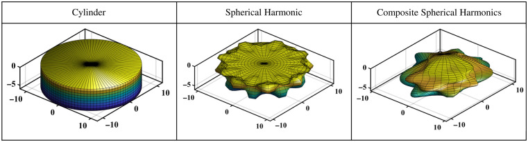
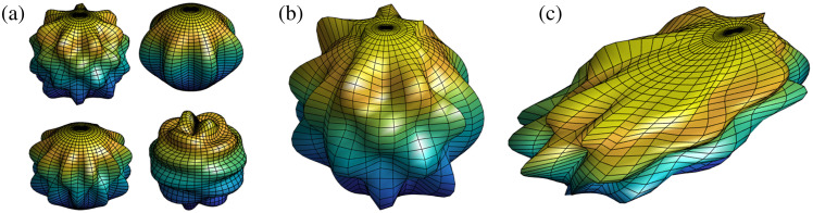
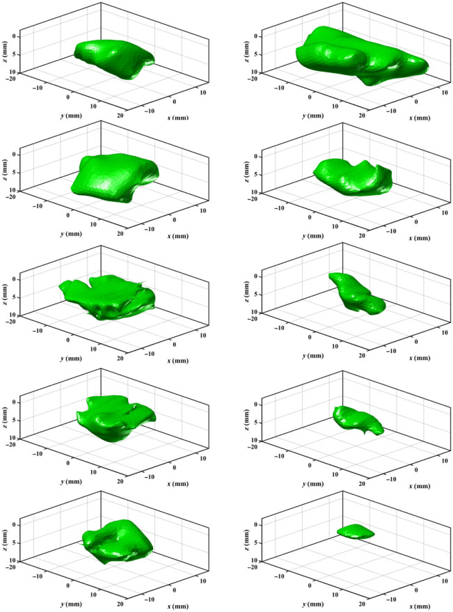
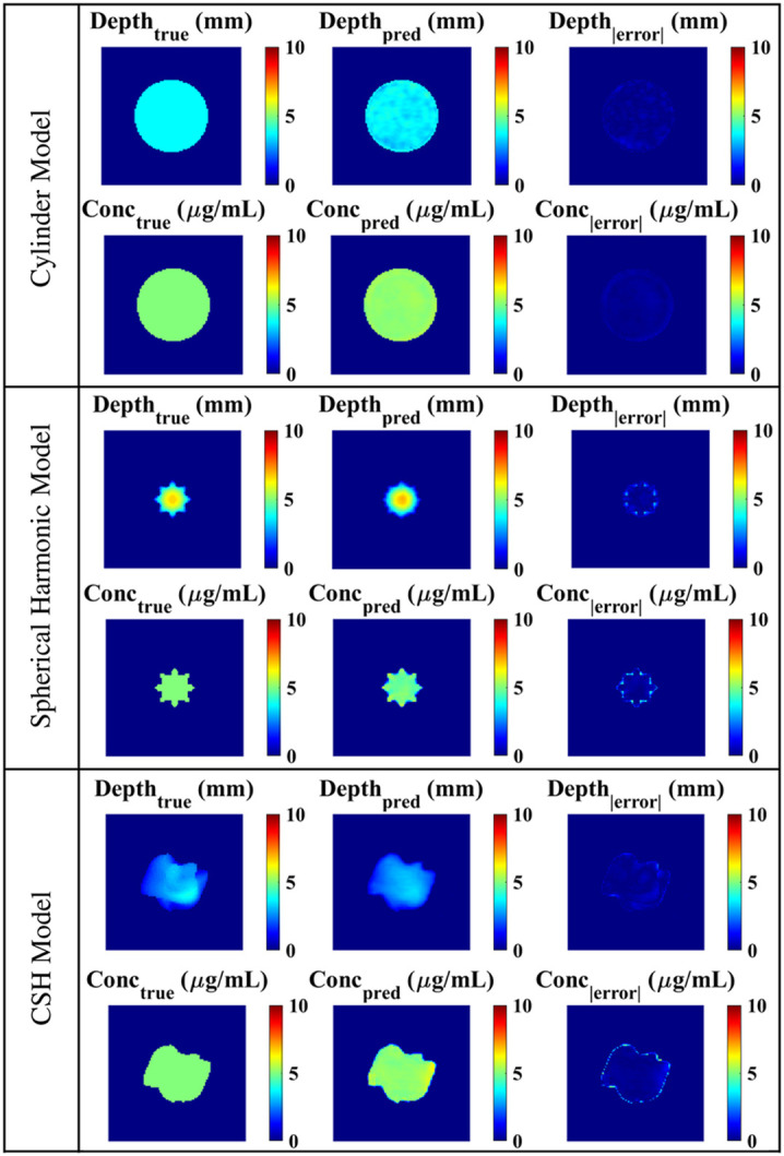
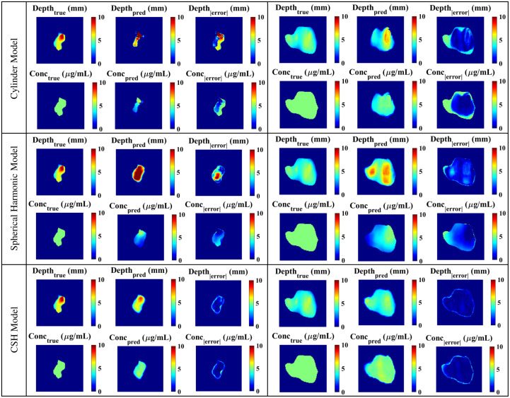
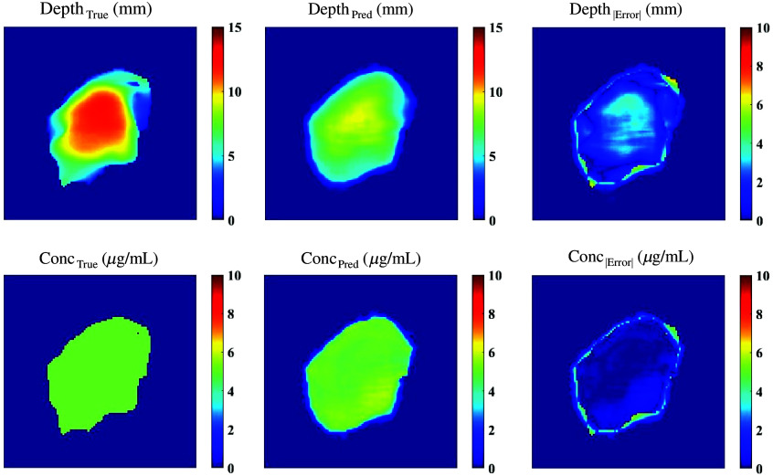
A convolutional neural network was designed to produce tumor depth and concentration maps from SFDI images. Three representations of oral cancer lesions were developed to train the DL architecture: cylinders, spherical harmonics, and composite spherical harmonics (CSHs). Each model was validated with SFDI images of patient-derived tongue tumors, and the CSH model was further validated with optical phantoms.
The performance of the CSH model was superior when presented with patient-derived tumors ( ). The CSH model could predict depth and concentration within 0.4 mm and , respectively, for tumors with depths less than 10 mm.
A DL-enabled SFDI system trained with CSH demonstrates promise in defining the deep margins of oral tumors.
口腔癌手术需要准确的边缘描绘,以平衡完全切除和术后功能。目前的荧光成像系统提供二维边缘评估,但在切除前无法量化肿瘤深度。利用结构光结合深度学习(DL)可能提供近实时的三维边缘检测。
开发了一种具有 DL 功能的荧光空间频域成像(SFDI)系统,该系统经过肿瘤模型训练,可量化口腔肿瘤的深度。
设计了一个卷积神经网络,可从 SFDI 图像中生成肿瘤深度和浓度图。开发了三种口腔癌病变的表示形式来训练 DL 架构:圆柱体、球谐函数和复合球谐函数(CSH)。每个模型都使用源自患者的舌肿瘤的 SFDI 图像进行了验证,CSH 模型还使用光学模型进行了验证。
当呈现源自患者的肿瘤时,CSH 模型的性能更优( )。CSH 模型可以预测深度和浓度,对于深度小于 10 毫米的肿瘤,分别在 0.4 毫米和 以内。
经过 CSH 训练的具有 DL 功能的 SFDI 系统在定义口腔肿瘤的深部边界方面具有潜力。