Carol Davila University of Medicine and Pharmacy, Bucharest, Romania.
Petroleum-Gas University, Ploiesti, Romania.
J Med Life. 2024 Jun;17(6):555-563. doi: 10.25122/jml-2024-0283.
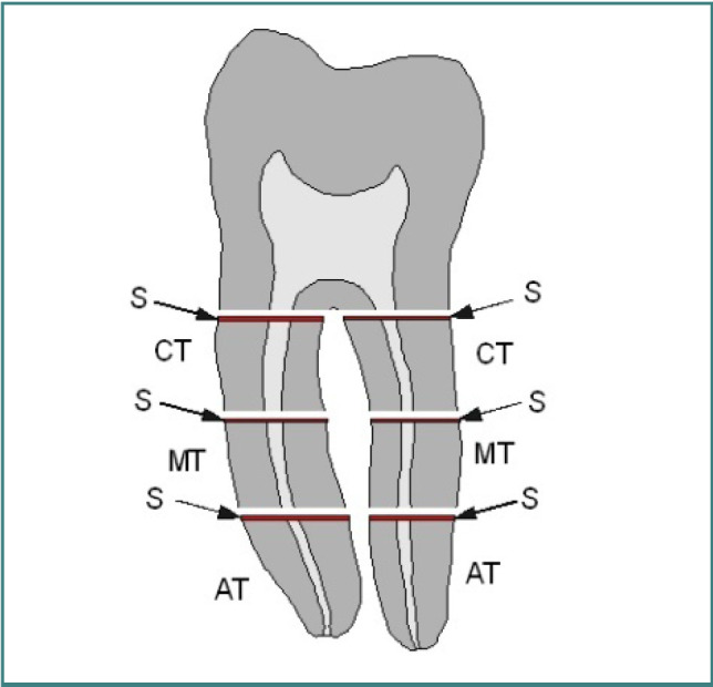
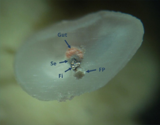
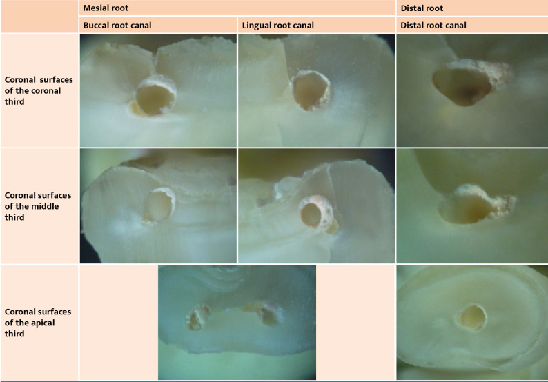
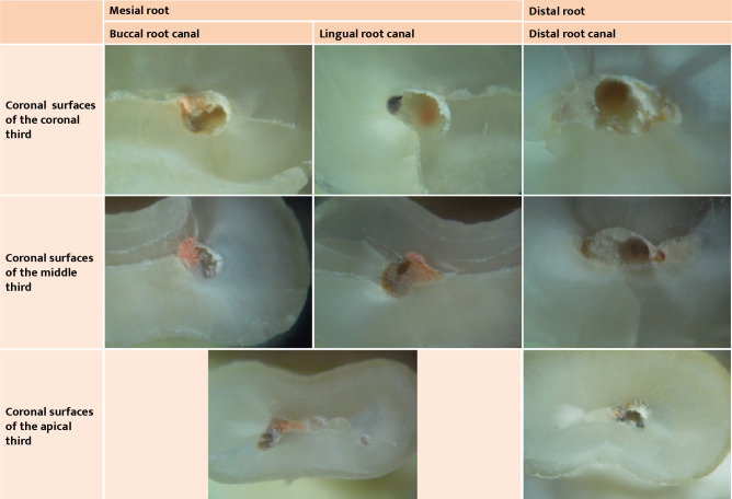
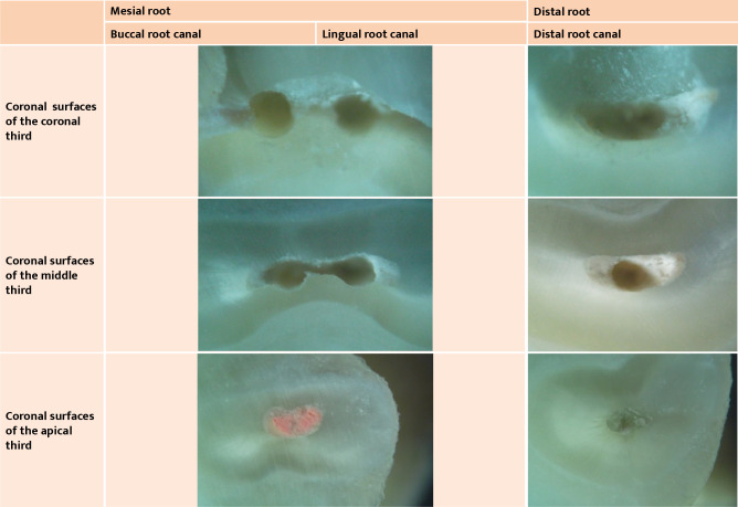
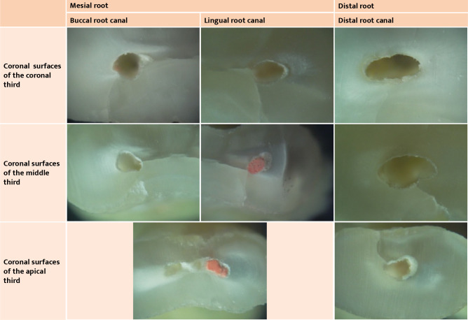
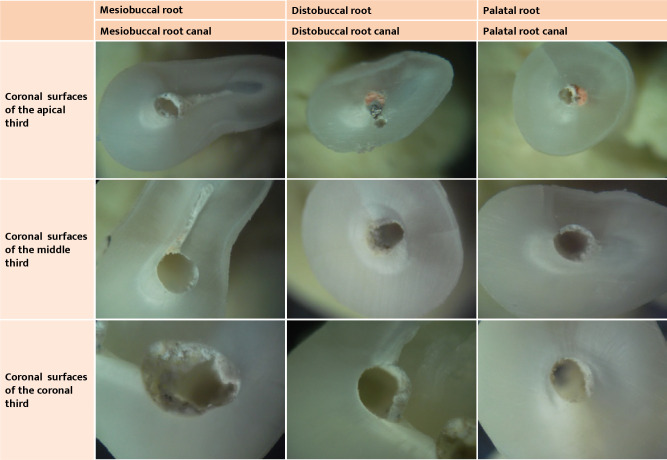
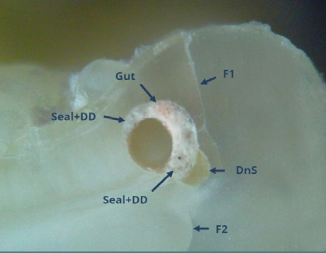
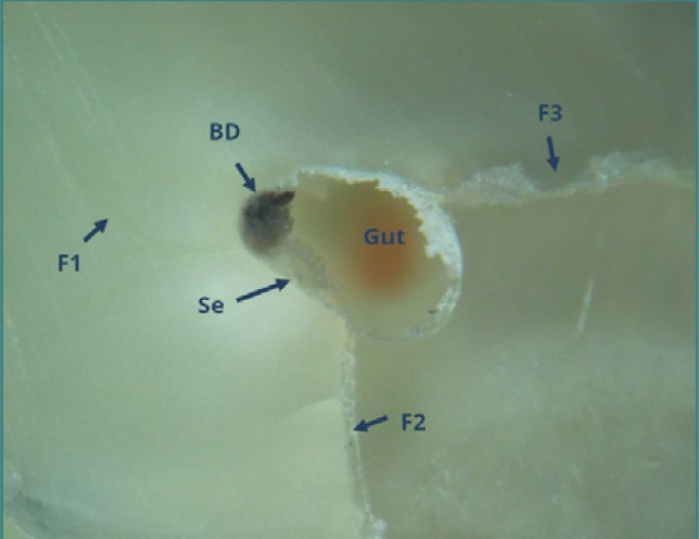
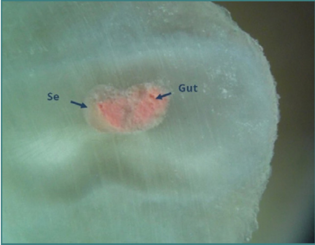
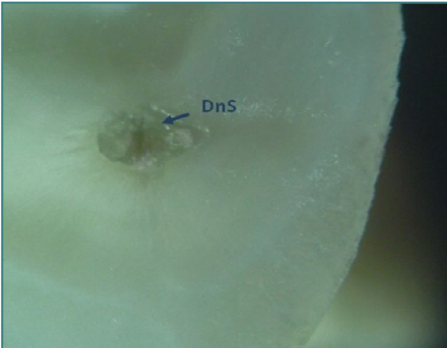
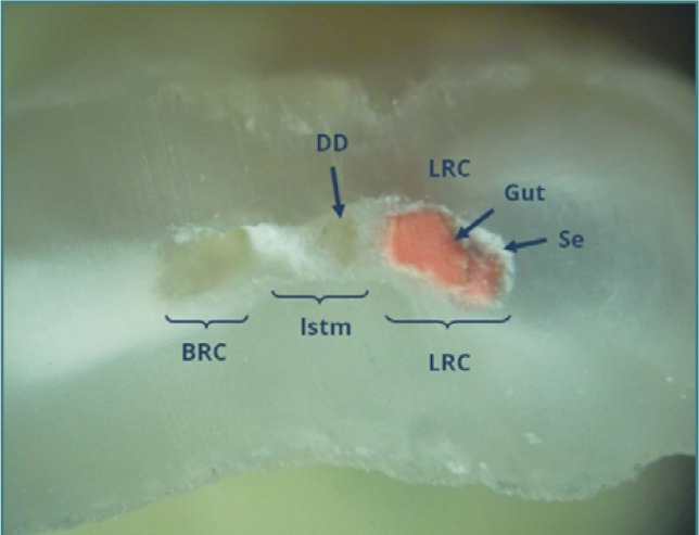
This study analyzed the effectiveness of root canal filling removal in lower molars performed by beginner operators using optical microscopy. A total of 55 mandibular first and second molars with mesial roots exhibiting an average curvature of 10-20° were selected based on preoperative radiographs. Instrumentation was done with ProTaper Gold (Dentsply Sirona) up to F2 (25/.08), using 2ml of 2.5% NaOCl irrigation solution after each file. Root canal obturation was performed using gutta-percha points with cold lateral condensation and Sealapex (Kerr Dental). Coronal fillings were made with composite resin and stored in distilled water for two years. Removal of the root canal fillings was performed with AF Retreatment Rotary (AFRR) and AF Blue R3 (AFBR3) (Fanta Dental Materials) under reciprocating motion with 2.5% NaOCl irrigation. Cross-sections of the coronal, middle, and apical thirds were analyzed at 40x magnification using a STEINDORFF POL microscope with a digital camera. Image analysis was conducted using Image J software, version 1.54, to determine the efficiency of root canal filling removal by percentage. Statistical analysis via one-way ANOVA revealed significant differences between distal and mesial roots ( < 0.05). Specifically, for mesial roots, the removal efficiency was 70.65% in the coronal third, 54.66% in the middle third, and 21.32% in the apical third. Significant difficulties were noted due to fractured files, calcifications, and debris accumulation in the isthmuses. The study concluded that the protocol using Fanta files demonstrated significant differences in removal efficiency correlated with root curvature, compounded by the inexperience of beginner operators. The findings highlight the challenges faced by novice practitioners in achieving effective root canal filling removal.
本研究通过光学显微镜分析了新手操作者在下颌磨牙中进行根管充填去除的效果。根据术前射线照片,选择了 55 颗下颌第一和第二磨牙,其近中根具有平均曲率为 10-20°。使用 ProTaper Gold(登士柏西诺德)器械进行根管预备,至 F2(25/.08),每次使用 2ml 2.5%次氯酸钠冲洗液冲洗。使用冷侧方加压技术(冷牙胶侧方加压技术)的牙胶尖进行根管充填,并用 Sealapex(科尔牙科)封闭。冠部填充物采用复合树脂,并在蒸馏水中储存两年。使用 AFRetreatment Rotary(AFRR)和 AF Blue R3(AFBR3)(Fanta Dental Materials)在根管内进行往复运动,同时使用 2.5%次氯酸钠冲洗液,去除根管内的填充物。使用 STEINDORFF POL 显微镜和数字相机在 40x 放大倍数下分析近中、中部和根尖三分之一的横断面。使用 Image J 软件(版本 1.54)进行图像分析,以确定根管填充物去除的效率。通过单因素方差分析进行统计分析,结果显示远中和近中根之间存在显著差异(<0.05)。具体而言,对于近中根,冠部三分之一的去除效率为 70.65%,中部三分之一为 54.66%,根尖三分之一为 21.32%。由于文件断裂、钙化和峡部碎屑堆积,发现了明显的困难。研究表明,使用 Fanta 文件的方案与根曲率相关,显示出显著的去除效率差异,这与新手操作者的经验不足有关。研究结果强调了新手从业者在实现有效根管填充去除方面面临的挑战。