Guerra Lucas Offenbecker, Leão Santos Ana Carolina, Cortinoz Janaina Rosa, Magalhães Renata Ferreira, Vasques Louise Idalgo, Leonardi Gislaine Ricci
Faculty of Medical Sciences, University of Campinas (UNICAMP), Campinas, Brazil.
ALS - Allergisa Pesquisa Dermato-Cosmetica Ltda., Campinas, Brazil.
Front Med (Lausanne). 2024 Sep 4;11:1391859. doi: 10.3389/fmed.2024.1391859. eCollection 2024.
Actinic keratosis (AK) is a highly prevalent pre-cancerous skin lesion that often leads to cutaneous squamous cell carcinoma. There are different stages of evolution of the disease and several features that characterize keratosis. This study aimed to develop a qualitative and quantitative visual diagnostic tool to facilitate the identification of the characteristics and severity of the main cellular attributes of AK and to show its applicability in evaluating the evolution or treatment through image analysis.
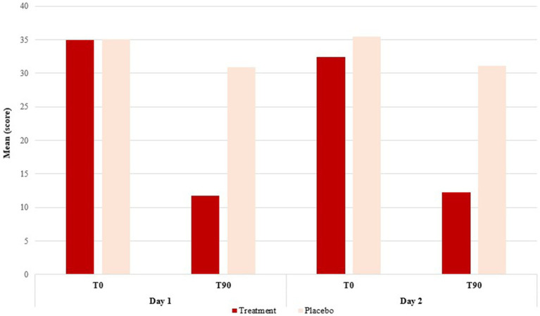
Literature research on the main scientific databases and in the institute's database was carried out to gather all the different levels of cellular transformation. To validate the scale, a preliminary characterization study was carried out with 21 subjects who had clinically diagnosed AK lesions to classify the attributes in each skin layer and test the accuracy of the diagnosis of the scale. Afterward, and to show the possibility of a follow-up with a topical treatment, the subjects were divided into two treatment groups, receiving either a cream formulation containing retinoic acid, or a placebo formula. The evaluation was carried out through confocal reflectance microscopy and a digital camera with dermoscopic quality before and after 90 days of treatment.
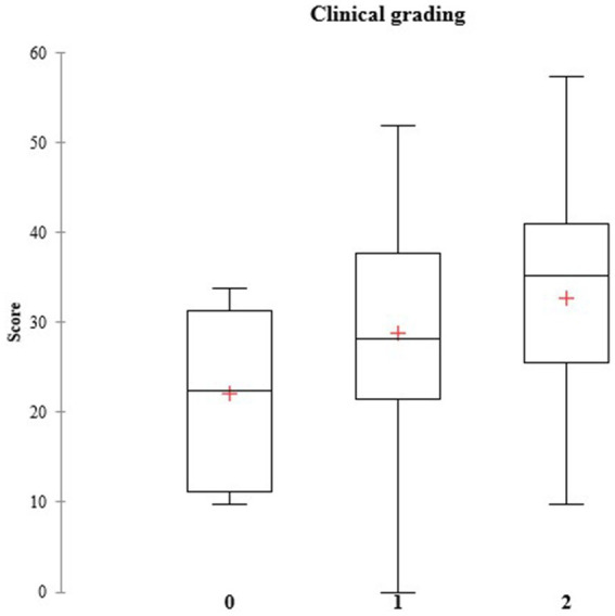
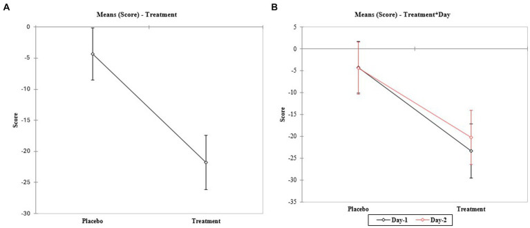
A table detailing the 18 attributes of AK, and a photographic scale containing RCM images graded by scores established for each characteristic and the frequency of spreading were developed. The results of the validation presented good repeatability, correlation with clinical evaluation, and capacity for differentiating treatments demonstrated by the significant improvement after topical treatment by the reduction of the score for 10 out of the 18 attributes. The preliminary study, evaluated by the detailed transformation scale highlights important differences in the subclinical approach that allows a deeper evaluation of the aspects of the lesion's re-incidence even after fully treated skin sites.
This study brings an innovative method based on RCM, to assist in the quantification of cell transformation level, provide early diagnosis, and deliver a powerful treatment evaluation tool to provide smoother treatment, as well as prevent re-incidence in the cases.
光化性角化病(AK)是一种高度常见的癌前皮肤病变,常发展为皮肤鳞状细胞癌。该疾病有不同的演变阶段和多种角化病特征。本研究旨在开发一种定性和定量的视觉诊断工具,以促进对AK主要细胞特征及严重程度的识别,并通过图像分析展示其在评估病情演变或治疗效果方面的适用性。
在主要科学数据库和该机构数据库中进行文献研究,以收集细胞转化的所有不同水平。为验证该量表,对21名临床诊断为AK病变的受试者进行了初步特征研究,以对各皮肤层的特征进行分类,并测试该量表诊断的准确性。之后,为展示局部治疗随访的可能性,将受试者分为两个治疗组,分别接受含维甲酸的乳膏制剂或安慰剂配方。在治疗90天前后,通过共聚焦反射显微镜和具有皮肤镜质量的数码相机进行评估。
制定了一个详细列出AK的18个特征的表格,以及一个包含通过为每个特征设定的分数分级的RCM图像和扩散频率的摄影量表。验证结果显示出良好的重复性、与临床评估的相关性,以及通过局部治疗后18个特征中的10个特征分数降低所证明的区分治疗效果的能力。通过详细转化量表评估的初步研究突出了亚临床方法中的重要差异,即使在皮肤部位完全治疗后,也能更深入地评估病变复发的各个方面。
本研究带来了一种基于RCM的创新方法,有助于量化细胞转化水平,提供早期诊断,并提供一个强大的治疗评估工具,以实现更平稳的治疗,并预防病例的复发。