Sidhu Paul S, Yusuf Gibran T, Sellars Maria E, Deganello Annamaria, Fang Cheng, Huang Dean Y H
King's College London, London, UK.
King's College Hospital, NHS Foundation Trust, London, UK.
Abdom Radiol (NY). 2025 Mar;50(3):1363-1375. doi: 10.1007/s00261-024-04587-z. Epub 2024 Sep 19.
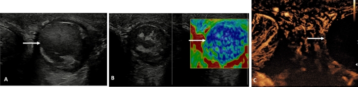
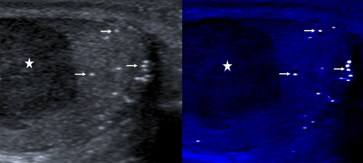
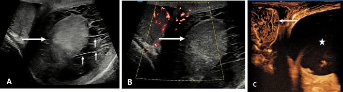
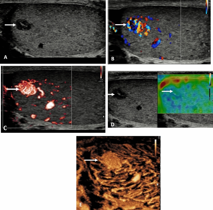
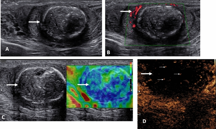
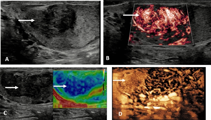
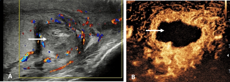
The innovative techniques in ultrasound have added a new dimension to investigating superficially located areas such as the contents of the scrotal sac. High frequency transducers, improved technology with the addition of elastography, contrast enhanced ultrasound and microvascular imaging has resulted in a further improvement in diagnostic capabilities. The ability to clearly demonstrate the presence or absence of vascularity within the area under investigation adds an additional dimension to operator confidence in establishing the presence of infarction, global or segmental, or the walls and cavity of an abscess in the testis or epididymis. Increased vascularity of a tumor aids the differential diagnosis based on the flow dynamics of the microbubble contrast, benign lesions likely to retain contrast. Elastography has the ability to ascertain the stiffness of tissue, and when used in conjunction with other ultrasound methods adds to the understanding of the likelihood of a malignant abnormality being present. All the different techniques come under the umbrella term 'multiparametric ultrasound', with the application in the scrotal sac detailed in this article.
超声领域的创新技术为诸如阴囊内容物等浅表部位的检查增添了新的维度。高频换能器、弹性成像、超声造影及微血管成像等技术的改进,进一步提升了诊断能力。清晰显示被检查区域有无血管的能力,为操作者判断梗死(整体或节段性)、睾丸或附睾脓肿的壁及腔隙的存在增添了信心。肿瘤血管增多有助于基于微泡造影剂的血流动力学进行鉴别诊断,良性病变可能会保留造影剂。弹性成像能够确定组织的硬度,与其他超声方法结合使用时,有助于加深对存在恶性异常可能性的理解。所有这些不同的技术都属于“多参数超声”这一统称,本文详细介绍了其在阴囊中的应用。