Jiang Xiao, Tivnan Matthew, Zhang Xiaoxuan, Stayman J Webster, Gang Grace J
Department of Biomedical Engineering, Johns Hopkins University, Baltimore MD, 21205, USA.
Department of Radiology, Harvard Medical School/Massachusetts General Hospital, Boston MA, 02114, USA.
Proc SPIE Int Soc Opt Eng. 2024 Feb;12925. doi: 10.1117/12.3006385. Epub 2024 Apr 1.
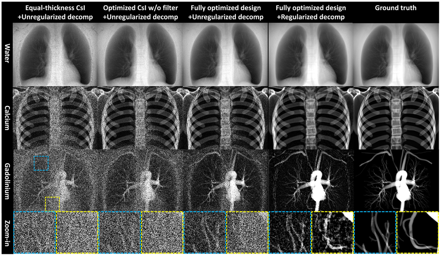
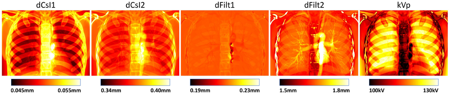
Spectral radiography and fluoroscopy with multi-layer flat-panel detectors (FPD) is being actively investigated in a range of clinical applications. For applications involving contrast administration, maximal contrast resolution is achieved when overlaying background anatomy is completely removed. This calls for three-material decomposition (soft tissue, bone, and contrast) enabled by measurements in three energy channels. We have previously demonstrated the feasibility of such decomposition using a triple-layer detector. While algorithmic solutions can be adopted to mitigate noise in the material basis images, in this work, we seek to fundamentally improve the conditioning of the problem through optimized system design. Design parameters include source voltage, the thickness of the top two CsI scintillators, and the thickness of two copper interstitial filters. The design objective is to minimize noise in the basis image containing contrast, chosen as gadolinium in this work to improve separation from bone. The optimized design was compared with other designs with unoptimized scintillator thickness and/or without interstitial filtration. Results show that CsI thickness optimization and interstitial filtration can significantly reduce noise in the gadolinium image by 35.7% and 42.7% respectively within a lung ROI, which in turn boosts detectability of small vessels. Gadolinium and bone signals are separated in all cases. Visualization of coronary vessels is enabled by the combining optimized system design and regularization. Results from this work demonstrate that three-material decomposition can be significantly improved with system design optimization. Optimized designs obtained from this work can inform imaging techniques selection and triple-layer detector fabrication for spectral radiography.
多层平板探测器(FPD)的光谱射线照相术和荧光透视术正在一系列临床应用中得到积极研究。对于涉及造影剂注入的应用,当完全去除重叠的背景解剖结构时,可实现最大对比度分辨率。这需要通过在三个能量通道进行测量来实现三材料分解(软组织、骨骼和造影剂)。我们之前已经证明了使用三层探测器进行这种分解的可行性。虽然可以采用算法解决方案来减轻材料基图像中的噪声,但在这项工作中,我们试图通过优化系统设计从根本上改善问题的条件。设计参数包括源电压、顶部两个碘化铯闪烁体的厚度以及两个铜间隙滤光片的厚度。设计目标是最小化包含造影剂的基图像中的噪声,在这项工作中选择钆以改善与骨骼的分离。将优化设计与闪烁体厚度未优化和/或没有间隙过滤的其他设计进行了比较。结果表明,碘化铯厚度优化和间隙过滤可分别在肺部感兴趣区域内将钆图像中的噪声显著降低35.7%和42.7%,这反过来提高了小血管的可检测性。在所有情况下,钆和骨骼信号都能分离。通过结合优化的系统设计和正则化,可以实现冠状动脉血管的可视化。这项工作的结果表明,通过系统设计优化可以显著改善三材料分解。从这项工作中获得的优化设计可为光谱射线照相术的成像技术选择和三层探测器制造提供参考。