Fujioka Kosuke, Katada Yoshiaki, Mataki Kentaro, Tsukanishi Toshinori, Ishii Tomoo, Morishita Yukio, Sugahara Shinji
Department of Radiology, Tokyo Medical University Ibaraki Medical Center, 3-20-1, Chuo, Inashiki-gun Ami-machi, Ibaraki 300-0395, Japan.
Department of Orthopedic Surgery, Tokyo Medical University Ibaraki Medical Center, 3-20-1, Chuo, Inashiki-gun Ami-machi, Ibaraki 300-0395, Japan.
Radiol Case Rep. 2024 Sep 10;19(12):5692-5695. doi: 10.1016/j.radcr.2024.08.042. eCollection 2024 Dec.
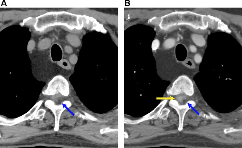
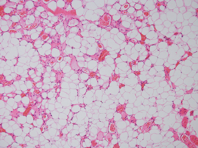
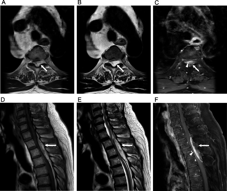
Spinal angiolipomas are rare benign tumors composed of mature adipose cells and blood vessel walls. We report the case of a patient with a spinal epidural angiolipoma who presented with paraplegia and was treated by urgent tumor resection and decompression. The patient was a 79-year-old man who presented to our hospital with a 6-month history of numbness in both lower limbs. Plain CT showed a tumor-like lesion with a predominantly fatty component on the dorsal epidural surface at the Th2-4 level, and contrast-enhanced CT showed a relatively strongly heterogeneously enhancing lesion. Gadolinium (Gd) -enhanced MRI also showed a well-defined spindle-shaped lesion measuring 2.4 × 1.0 × 6.5 cm in size that was visualized as a heterogeneous high signal intensity on both T1- and T2-weighted images and showed strong heterogeneous enhancement on fat-saturated Gd-enhanced T1-weighted images. We performed Th1-4 laminectomy and tumor resection and the patient was discharged home, with no numbness in the lower limbs.
脊髓血管脂肪瘤是一种罕见的由成熟脂肪细胞和血管壁组成的良性肿瘤。我们报告了一例患有脊髓硬膜外血管脂肪瘤的患者,该患者出现截瘫,并接受了紧急肿瘤切除和减压治疗。患者为一名79岁男性,因双下肢麻木6个月前来我院就诊。平扫CT显示在胸2 - 4水平的硬膜背侧表面有一个以脂肪成分为主的肿瘤样病变,增强CT显示为一个强化相对较强的不均匀强化病变。钆(Gd)增强MRI也显示一个边界清晰的梭形病变,大小为2.4×1.0×6.5 cm,在T1加权和T2加权图像上均表现为不均匀高信号强度,在脂肪饱和Gd增强T1加权图像上显示出强烈的不均匀强化。我们进行了胸1 - 4椎板切除术和肿瘤切除术,患者出院回家时双下肢无麻木感。