Wells Simon P, O'Shea Christopher, Hayes Sarah, Weeks Kate L, Kirchhof Paulus, Delbridge Lea M D, Pavlovic Davor, Bell James R
Department of Anatomy and Physiology, University of Melbourne, Parkville, Victoria, Australia.
Institute of Cardiovascular Sciences, University of Birmingham, Birmingham, United Kingdom.
J Mol Cell Cardiol Plus. 2024 Sep;9:100079. doi: 10.1016/j.jmccpl.2024.100079.
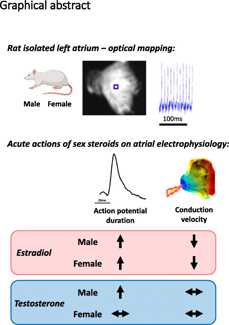
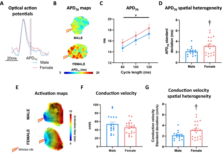
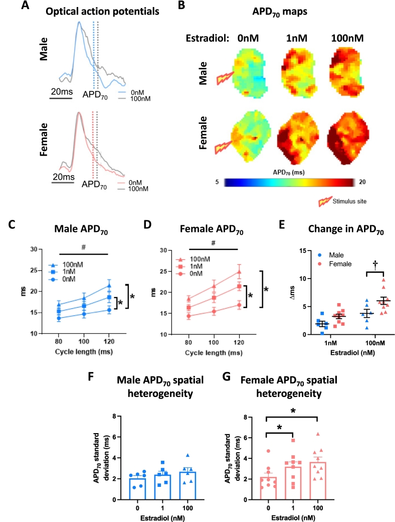
The electrophysiological properties of the hearts of women and men are different. These differences are at least partly mediated by the actions of circulating estrogens and androgens on the cardiomyocytes. Experimentally, much of our understanding in this field is based on studies focusing on ventricular tissue, with considerably less known in the context of atrial electrophysiology. The aim of this investigation was to compare the electrophysiological properties of male and female atria and assess responses to acute sex steroid exposure. Age-matched adult male and female C57BL/6 mice were anesthetized (4 % isoflurane) and left atria isolated. Atria were loaded with Di-4-ANEPPS voltage sensitive dye and optical mapping performed to assess action potential duration (APD; at 10 %, 20 %, 30 %, 50 %, and 70 % repolarization) and conduction velocity in the presence of 1 nM and 100 nM 17β-estradiol or testosterone. Male and female left atria demonstrated similar baseline action potential duration and conduction velocity, with significantly greater APD spatial heterogeneity evident in females. 17β-estradiol prolonged action potential duration in both sexes - an effect that was augmented in females. Atrial conduction was slowed in the presence of 100 nM 17β-estradiol in both males and females. Testosterone prolonged action potential duration in males only and did not modulate conduction velocity in either sex. This study provides novel insights into male and female atrial electrophysiology and its regulation by sex steroids. As systemic sex steroid levels change and intra-cardiac estrogen synthesis capacity increases with aging, these actions may have an increasingly important role in determining atrial arrhythmia vulnerability.
男性和女性心脏的电生理特性有所不同。这些差异至少部分是由循环中的雌激素和雄激素对心肌细胞的作用介导的。在实验中,我们在该领域的大部分理解都基于对心室组织的研究,而关于心房电生理学的了解则少得多。本研究的目的是比较雄性和雌性心房的电生理特性,并评估对急性性类固醇暴露的反应。将年龄匹配的成年雄性和雌性C57BL/6小鼠麻醉(4%异氟烷),分离出左心房。将心房加载Di-4-ANEPPS电压敏感染料,并进行光学映射以评估在存在1 nM和100 nM 17β-雌二醇或睾酮的情况下的动作电位持续时间(APD;在复极化10%、20%、30%、50%和70%时)和传导速度。雄性和雌性左心房表现出相似的基线动作电位持续时间和传导速度,雌性中APD的空间异质性明显更大。17β-雌二醇使两性的动作电位持续时间延长——这种作用在雌性中增强。在雄性和雌性中,100 nM 17β-雌二醇存在时心房传导减慢。睾酮仅使雄性的动作电位持续时间延长,对两性的传导速度均无调节作用。本研究为雄性和雌性心房电生理学及其受性类固醇调节提供了新的见解。随着全身性激素水平的变化以及心脏内雌激素合成能力随年龄增长而增加,这些作用在决定心房心律失常易感性方面可能发挥越来越重要的作用。