Kurihara Masanori, Arakawa Akira, Tokumaru Aya Midori, Matsubara Tomoyasu, Eguchi Hiroto, Shimo Yasushi, Hasegawa Masato, Kanemaru Kazutomi, Takeda Katsuhiko, Iwata Atsushi, Murayama Shigeo, Saito Yuko
Department of Neurology, Tokyo Metropolitan Institute for Geriatrics and Gerontology, Tokyo, Japan.
Department of Neuropathology (Brain Bank for Aging Research), Tokyo Metropolitan Institute for Geriatrics and Gerontology, Tokyo, Japan.
eNeurologicalSci. 2024 Sep 4;37:100526. doi: 10.1016/j.ensci.2024.100526. eCollection 2024 Dec.
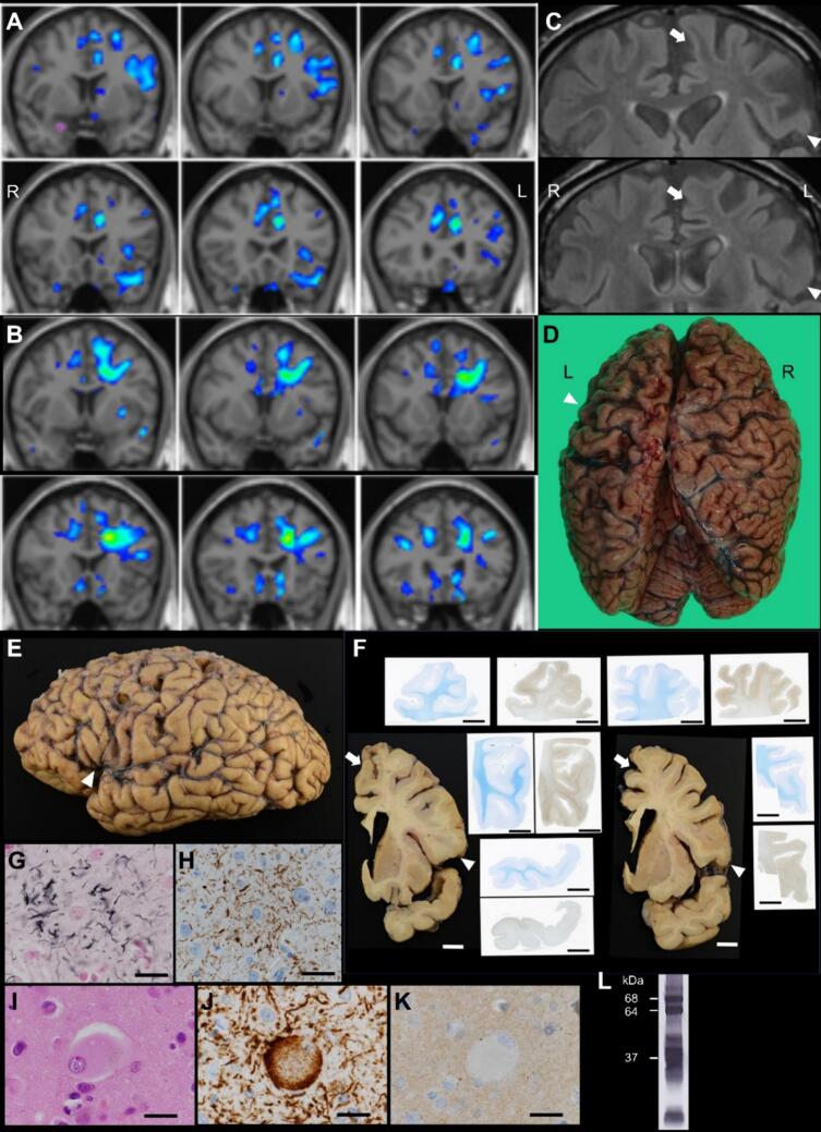
A 72-year-old man presented with a 6-month history of decreased voluntary speech. Sparse speech and decreased word fluency were observed. Articulation, naming, comprehension, and repetition were preserved. Agrammatism and paraphasia were not observed. These characteristics matched those reported as dynamic aphasia. Other findings were mild behavioral symptoms, recent memory impairment, and right hemiparkinsonism. The patient's voluntary speech continued to reduce and behavioral symptoms progressed. Brain MRI including voxel-based morphometric analysis showed left-dominant white matter volume reduction in the frontal lobe including those between the left supplementary motor area (SMA)/preSMA and the frontal operculum, likely involving the frontal aslant tract (FAT). The patient became completely mute after two years from disease onset and died of aspiration pneumonia. The neuropathological diagnosis was corticobasal degeneration (CBD). This case suggests that dynamic aphasia may be the initial sign of CBD and that early involvement of left FAT may be responsible for this feature.
一名72岁男性,有6个月的自主言语减少病史。观察到言语稀疏且词汇流畅性降低。发音、命名、理解和复述功能保留。未观察到语法缺失和言语错乱。这些特征与报道的动态性失语相符。其他发现包括轻度行为症状、近期记忆障碍和右侧偏侧帕金森综合征。患者的自主言语持续减少,行为症状进展。包括基于体素的形态学分析在内的脑部MRI显示,左侧额叶白质体积减少,包括左侧辅助运动区(SMA)/前辅助运动区与额盖之间的区域,可能累及额斜束(FAT)。发病两年后患者完全失语,死于吸入性肺炎。神经病理学诊断为皮质基底节变性(CBD)。该病例提示动态性失语可能是CBD的初始症状,左侧FAT的早期受累可能是这一特征的原因。