Walha Yasmin, Rekik Mona, Moalla Khadija Sonda, Kammoun Sonda, Ayadi Omar, Mhiri Chokri, Dammak Mariem, Trigui Amira
Department of Ophthalmology, Habib Bourguiba Hospital, 3029 Sfax, Tunisia.
Department of Neurology, Habib Bourguiba Hospital, 3029 Sfax, Tunisia.
eNeurologicalSci. 2024 Sep 4;37:100525. doi: 10.1016/j.ensci.2024.100525. eCollection 2024 Dec.
To analyze changes in peripapillary retinal nerve fiber layer (RNFL) and ganglion cell complex (GCC) thickness in migraine patients with and without aura compared to healthy controls and to identify factors influencing the occurrence of these anomalies.
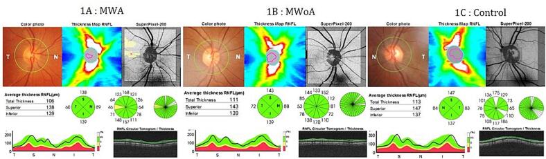
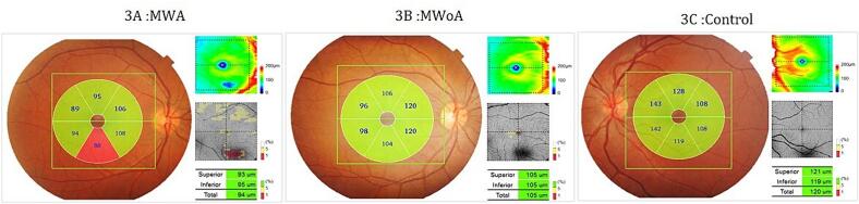
This is a cross-sectional case-control study including migraine patients and control subjects. All patients and controls underwent a complete ophthalmological examination, RNFL and GCC thickness measurements using a spectral domain-OCT device.The duration of migraine, the frequency and duration of migraine attacks, the migraine disability assessment (MIDAS) and migraine severity scale (MIGSEV) questionnaire scores were recorded.
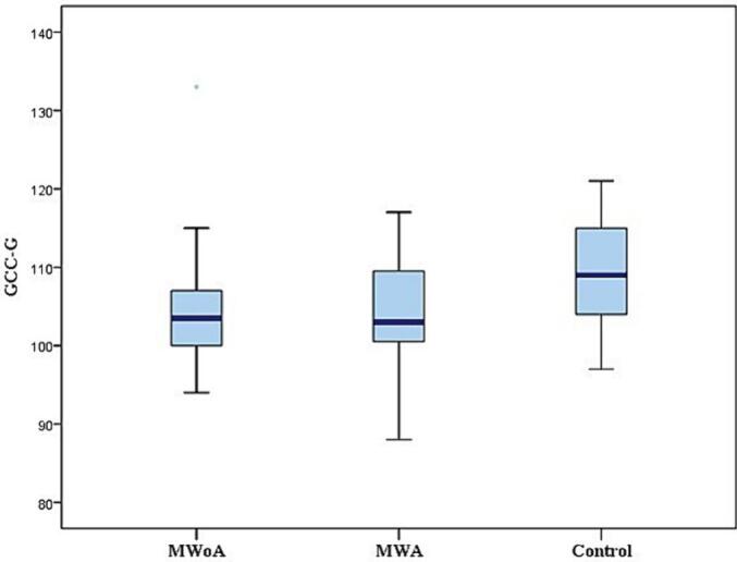
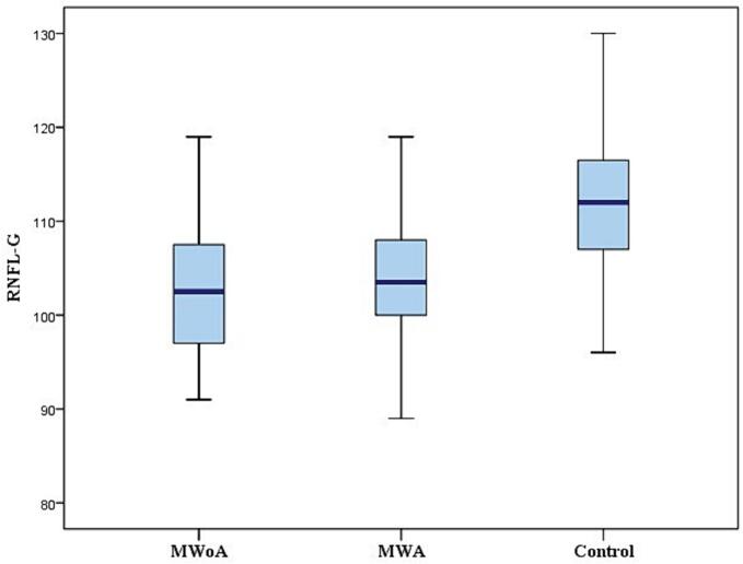
One hundred and twenty eyes from 60 patients (60 eyes in the migraine without aura (MWoA) group and 60 eyes in the migraine with aura (MWA) group) were included. Control group included 30 age and gender matched healthy participants (60 eyes). OCT revealed that RNFL and GCC thickness were significantly reduced in the migraine without aura (MWoA) and in the migraine with aura (MWA) groups compared to the control group and in the migraine with aura (MWA) group compared to the migraine without aura (MWoA) group. Prolonged disease duration was associated to decreased GCC thickness. RNFL and GCC thickness were correlated to disease severity, attack frequency and duration. In the multivariate study, duration of migraine and attack frequency were the main determinant factors of nasal GCC thickness. Disease severity was the main determinant of RNFL and GCC thickness, with the exception of the nasal sector.
Our study emphasize the significant impact of both types of migraine on retinal structures. OCT would serve as a valuable biomarker in migraine.
分析有先兆和无先兆偏头痛患者与健康对照者相比,视盘周围视网膜神经纤维层(RNFL)和神经节细胞复合体(GCC)厚度的变化,并确定影响这些异常发生的因素。
这是一项横断面病例对照研究,纳入了偏头痛患者和对照者。所有患者和对照者均接受了全面的眼科检查,使用光谱域光学相干断层扫描(OCT)设备测量RNFL和GCC厚度。记录偏头痛的病程、偏头痛发作的频率和持续时间、偏头痛残疾评估(MIDAS)和偏头痛严重程度量表(MIGSEV)问卷得分。
纳入了60例患者的120只眼(无先兆偏头痛(MWoA)组60只眼,有先兆偏头痛(MWA)组60只眼)。对照组包括30名年龄和性别匹配的健康参与者(60只眼)。OCT显示,与对照组相比,无先兆偏头痛(MWoA)组和有先兆偏头痛(MWA)组以及有先兆偏头痛(MWA)组与无先兆偏头痛(MWoA)组相比,RNFL和GCC厚度均显著降低。病程延长与GCC厚度降低相关。RNFL和GCC厚度与疾病严重程度、发作频率和持续时间相关。在多变量研究中,偏头痛病程和发作频率是鼻侧GCC厚度的主要决定因素。除鼻侧扇形区外,疾病严重程度是RNFL和GCC厚度的主要决定因素。
我们的研究强调了两种类型偏头痛对视网膜结构的显著影响。OCT可作为偏头痛中有价值的生物标志物。