Li Yang, Yang Li, Gu Xiaolong, Wang Xiangming, Wang Qi, Shi Gaofeng, Zhang Andu, Deng Huiyan, Zhao Xiaopeng, Ren Jialiang, Miao Aijun, Li Shaolian
The Fourth Hospital of Hebei Medical University, Shijiazhuang, China.
GE Healthcare China, Beijing, China.
Abdom Radiol (NY). 2025 Apr;50(4):1475-1487. doi: 10.1007/s00261-024-04562-8. Epub 2024 Sep 23.
This study aimed to investigate whether contrast-enhanced computed tomography (CECT) based radiomics analysis could noninvasively predict the perineural invasion (PNI) in esophageal squamous cell carcinoma (ESCC).
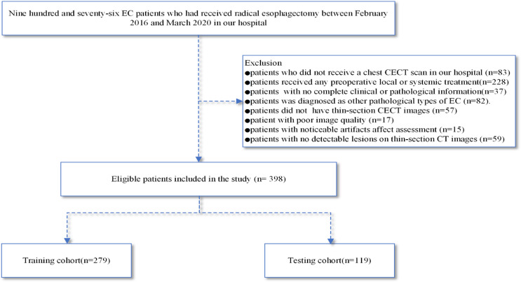
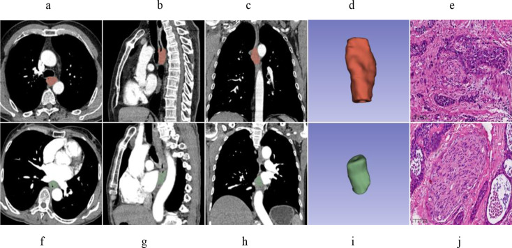
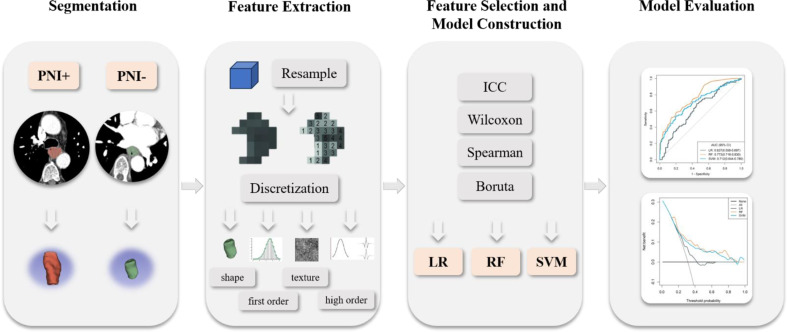
398 patients with ESCC who underwent resection between February 2016 and March 2020 were retrospectively enrolled in this study. Patients were randomly divided into training and testing cohorts in a 7:3 ratio. Radiomics analysis was performed on the arterial phase images of CECT scans. From these images, 1595 radiomics features were initially extracted. The intraclass correlation coefficient (ICC), wilcoxon rank-sum test, spearman correlation analysis, and boruta algorithm were used for feature selection. Logistic regression (LR), random forest (RF), and support vector machine (SVM) models were established to preidict the PNI status. The performance of these radiomics models was assessed by the area under the receiver operating characteristic curve (AUC). Decision curve analysis (DCA) was conducted to evaluate their clinical utility.
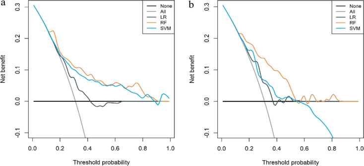
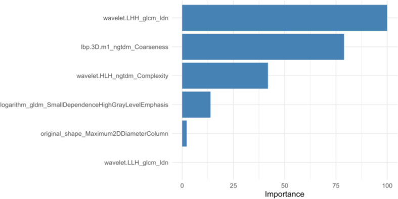
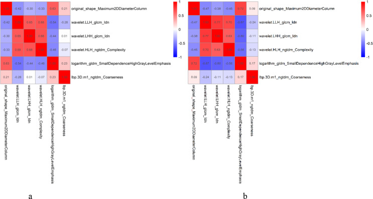
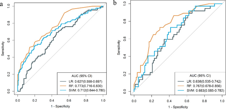
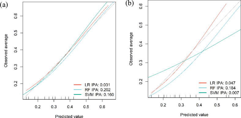
Six radiomics features were retained to build the radiomics models. Among these models, the random forest (RF) model demonstrated superior performance. In the training cohort, the AUC value of the RF model was 0.773, compared to 0.627 for the logistic regression (LR) model and 0.712 for the support vector machine (SVM) model. Similarly, in the testing cohort, the RF model achieved an AUC value of 0.767, outperforming the LR model at 0.638 and the SVM model at 0.683. Decision curve analysis (DCA) suggested that the RF radiomics model exhibited the highest clinical utility.
CECT-based radiomics analysis, particularly utilizing the RF, can noninvasively predict the PNI in ESCC preoperatively. This novel approach could enhance patient management by providing personalized information, thereby facilitating the development of individualized treatment strategies for ESCC patients.
本研究旨在探讨基于对比增强计算机断层扫描(CECT)的影像组学分析能否无创预测食管鳞状细胞癌(ESCC)的神经周围侵犯(PNI)。
回顾性纳入2016年2月至2020年3月间接受手术切除的398例ESCC患者。患者按7:3的比例随机分为训练组和测试组。对CECT扫描的动脉期图像进行影像组学分析。从这些图像中,最初提取了1595个影像组学特征。采用组内相关系数(ICC)、威尔科克森秩和检验、斯皮尔曼相关分析和博鲁塔算法进行特征选择。建立逻辑回归(LR)、随机森林(RF)和支持向量机(SVM)模型来预测PNI状态。通过受试者操作特征曲线(AUC)下面积评估这些影像组学模型的性能。进行决策曲线分析(DCA)以评估其临床实用性。
保留6个影像组学特征来构建影像组学模型。在这些模型中,随机森林(RF)模型表现出卓越性能。在训练组中,RF模型的AUC值为0.773,而逻辑回归(LR)模型为0.627,支持向量机(SVM)模型为0.712。同样,在测试组中,RF模型的AUC值为0.767,优于LR模型的0.638和SVM模型的0.683。决策曲线分析(DCA)表明RF影像组学模型具有最高的临床实用性。
基于CECT的影像组学分析,尤其是利用RF,能够在术前无创预测ESCC中的PNI。这种新方法可通过提供个性化信息来加强患者管理,从而促进ESCC患者个体化治疗策略的制定。