Moreno-Gonzalez Mar, Hampton Katherine, Ruiz Paula, Beasy Gemma, Nagies Falk Sp, Parker Aimee, Lazenby James, Bone Caitlin, Alava-Arteaga Ane, Patel Meha, Hellmich Charlotte, Luri-Martin Pablo, Silan Ece, Philo Mark, Baker David, Rushbrook Simon M, Hildebrand Falk, Rushworth Stuart A, Beraza Naiara
Gut Microbes and Health Institute Strategic Programme, Quadram Institute Bioscience, Norwich Research Park, Norwich, UK.
Food, Microbiome and Health Institute Strategic Programme, Quadram Institute Bioscience, Norwich Research Park, Norwich, UK.
JHEP Rep. 2024 Jun 29;6(10):101159. doi: 10.1016/j.jhepr.2024.101159. eCollection 2024 Oct.
BACKGROUND & AIMS: Senescence has been reported to have differential functions in cholangiocytes and hepatic stellate cells (HSCs) during human and murine cholestatic disease, being detrimental in biliary cells and anti-fibrotic in HSCs. Cholestatic liver disease is associated with loss of intestinal barrier function and changes in the microbiome, the mechanistic cause of which is undetermined.
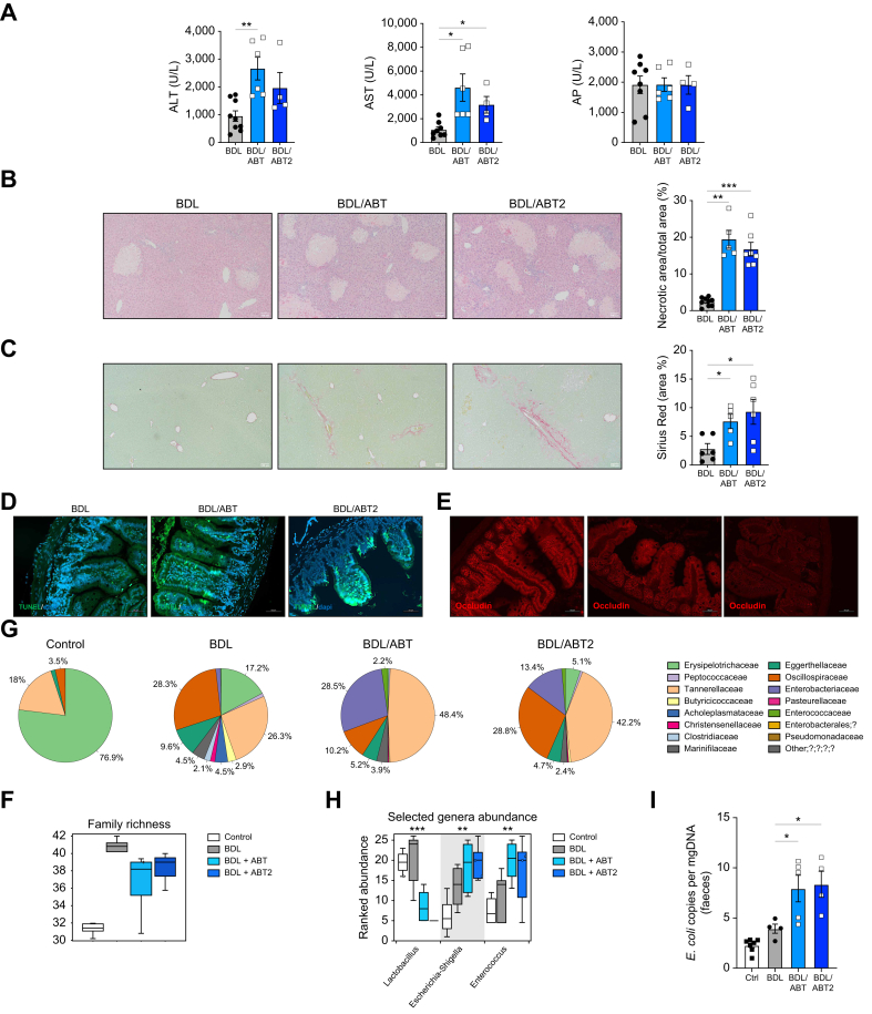
Intestinal samples were analysed from controls and patients with primary sclerosing cholangitis, as well as wild-type (WT) and p16-3MR transgenic mice. Cholestatic liver disease was induced by bile duct ligation (BDL) and DDC diet feeding. Fexaramine was used as an intestinal-restricted FXR agonist and antibiotics were given to eliminate the intestinal microbiome. Senescent cells were eliminated in p16-3MR mice with ganciclovir and in WT mice with the senolytic drug ABT-263. studies were done in intestinal CaCo-2 cells and organoids were generated from intestinal crypts isolated from mice.
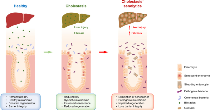
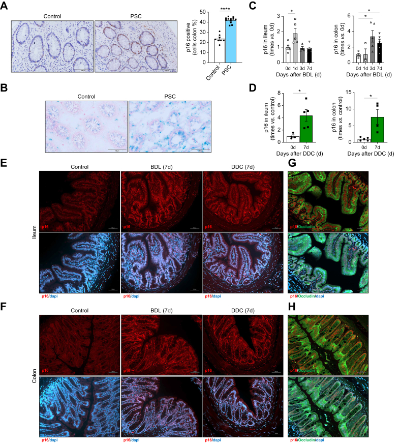
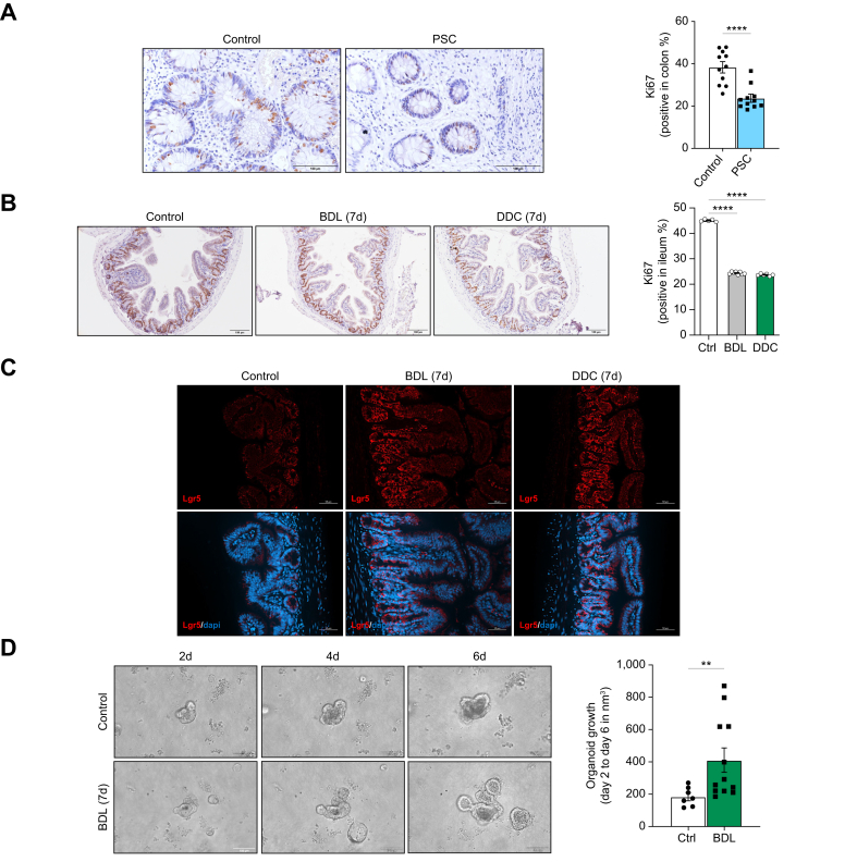
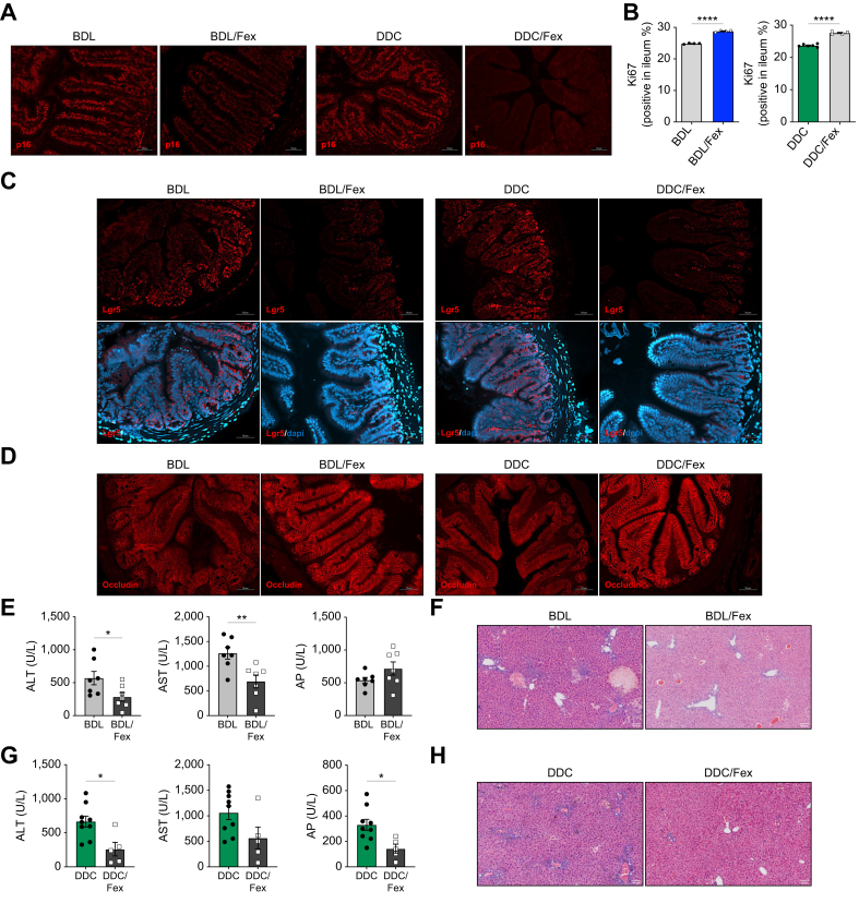
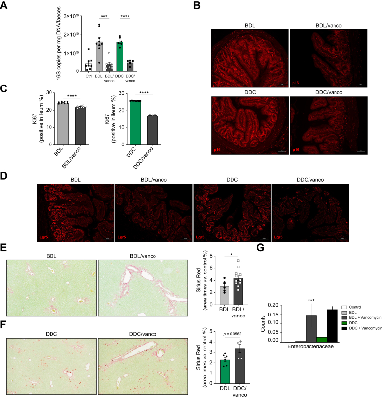
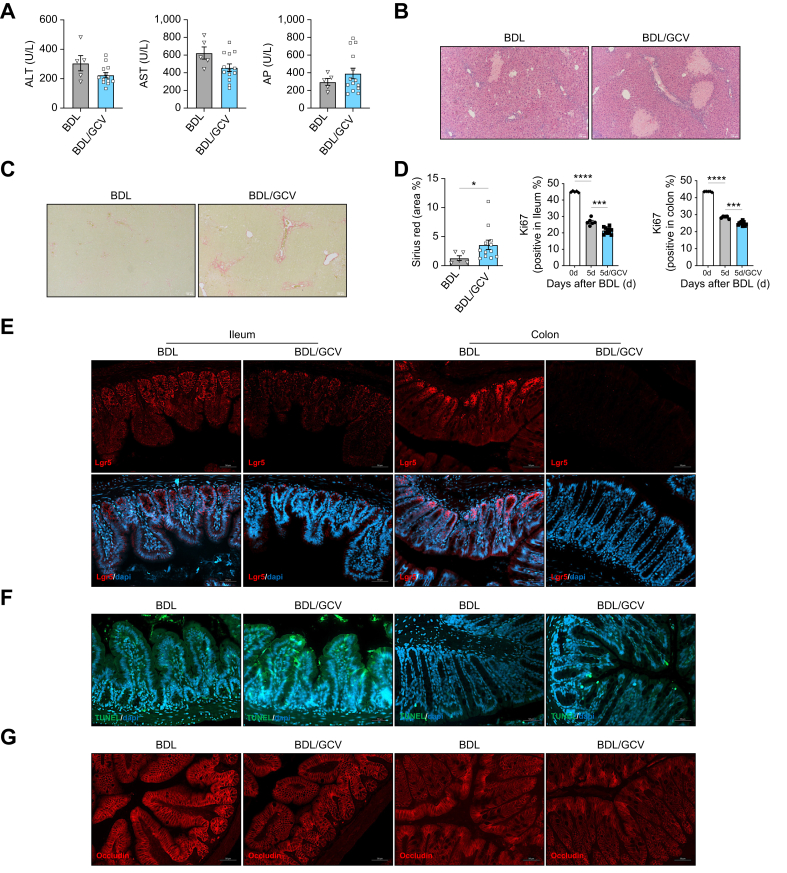
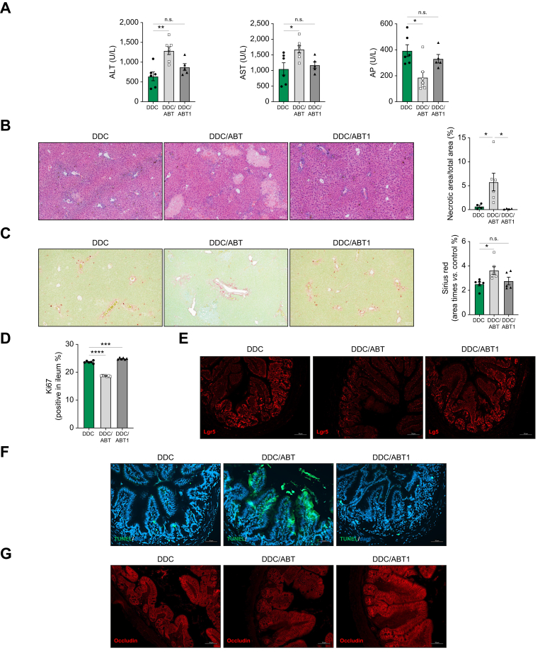
Herein, we show increased senescence in intestinal epithelial cells (IECs) in patients with primary sclerosing cholangitis and in mice after BDL and DDC diet feeding. Intestinal senescence was increased in response to reduced exposure to bile acids and increased presence of lipopolysaccharide and during cholestatic liver disease. Senescence of IECs was associated with lower proliferation but increased intestinal stem cell activation, as supported by increased organoid growth from intestinal stem cells. Elimination of senescent cells with genetic and pharmacological approaches exacerbated liver injury and fibrosis during cholestatic liver disease, which was associated with increased IEC apoptosis and permeability.
Senescence occurs in IECs during cholestatic disease and the elimination of senescent cells has a detrimental impact on the gut-liver axis. Our results point to cell-specific rather than systemic targeting of senescence as a therapeutic approach to treat cholestatic liver disease.
Cholestatic liver disease associates with the dysregulation of intestinal barrier function, while the mechanisms mediating the disruption of the gut-liver axis remain largely undefined. Here, we demonstrate that senescence, a cellular response to stress, is activated in intestinal cells during cholestatic liver disease in humans and mice. Mechanistically, we demonstrate that the reduction of bile acids and the increased presence of bacterial products mediate the activation of intestinal senescence during cholestatic liver disease. Importantly, the elimination of these senescent cells promotes further damage to the intestine that aggravates liver disease, with increased tissue damage and fibrosis. Our results provide evidence that therapeutic strategies to treat cholestatic liver disease by eliminating senescent cells may have unwanted effects in the intestine and support the need to develop cell/organ-specific approaches.
据报道,在人类和小鼠胆汁淤积性疾病中,衰老在胆管细胞和肝星状细胞(HSC)中具有不同的功能,对胆管细胞有害,而对HSC具有抗纤维化作用。胆汁淤积性肝病与肠道屏障功能丧失和微生物群变化有关,其机制尚不清楚。
分析了对照组和原发性硬化性胆管炎患者以及野生型(WT)和p16-3MR转基因小鼠的肠道样本。通过胆管结扎(BDL)和给予二氯二苯醚菊酯(DDC)饮食诱导胆汁淤积性肝病。使用非瑟酮胺作为肠道特异性法尼醇X受体(FXR)激动剂,并给予抗生素以消除肠道微生物群。用更昔洛韦在p16-3MR小鼠中清除衰老细胞,并用衰老溶解药物ABT-263在WT小鼠中清除衰老细胞。在肠道CaCo-2细胞中进行研究,并从小鼠分离的肠道隐窝中生成类器官。
在此,我们显示原发性硬化性胆管炎患者以及BDL和DDC饮食喂养后的小鼠肠道上皮细胞(IEC)中的衰老增加。在胆汁淤积性肝病期间,肠道衰老因胆汁酸暴露减少和脂多糖存在增加而增加。IEC衰老与较低的增殖相关,但肠道干细胞活化增加,这得到了肠道干细胞类器官生长增加的支持。通过遗传和药理学方法消除衰老细胞会加剧胆汁淤积性肝病期间的肝损伤和纤维化,这与IEC凋亡和通透性增加有关。
在胆汁淤积性疾病期间,IEC中会发生衰老,消除衰老细胞会对肠-肝轴产生有害影响。我们的结果表明,将衰老作为治疗胆汁淤积性肝病的治疗方法应针对细胞特异性而非全身性靶向。
胆汁淤积性肝病与肠道屏障功能失调有关,而介导肠-肝轴破坏的机制在很大程度上仍不清楚。在此,我们证明,衰老作为一种细胞应激反应,在人类和小鼠胆汁淤积性肝病期间在肠道细胞中被激活。从机制上讲,我们证明胆汁酸减少和细菌产物存在增加介导了胆汁淤积性肝病期间肠道衰老的激活。重要的是,消除这些衰老细胞会促进肠道进一步损伤,加剧肝病,增加组织损伤和纤维化。我们的结果提供了证据,表明通过消除衰老细胞来治疗胆汁淤积性肝病的治疗策略可能会对肠道产生不良影响,并支持开发细胞/器官特异性方法的必要性。