Alsuraih Mohammed, O'Hara Steven P, Woodrum Julie E, Pirius Nicholas E, LaRusso Nicholas F
Department of Biochemistry and Molecular Biology, Mayo Clinic, Rochester, MN, 55905, USA.
Mayo Clinic Graduate School of Biomedical Sciences, Mayo Clinic, Rochester, MN, 55905, USA.
JHEP Rep. 2021 Jan 27;3(3):100250. doi: 10.1016/j.jhepr.2021.100250. eCollection 2021 Jun.
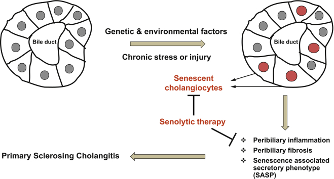
BACKGROUND & AIMS: Cholangiocyte senescence is important in the pathogenesis of primary sclerosing cholangitis (PSC). We found that CDKN2A (p16), a cyclin-dependent kinase inhibitor and mediator of senescence, was increased in cholangiocytes of patients with PSC and from a PSC mouse model (multidrug resistance 2; ). Given that recent data suggest that a reduction of senescent cells is beneficial in different diseases, we hypothesised that inhibition of cholangiocyte senescence would ameliorate disease in mice.
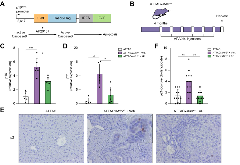
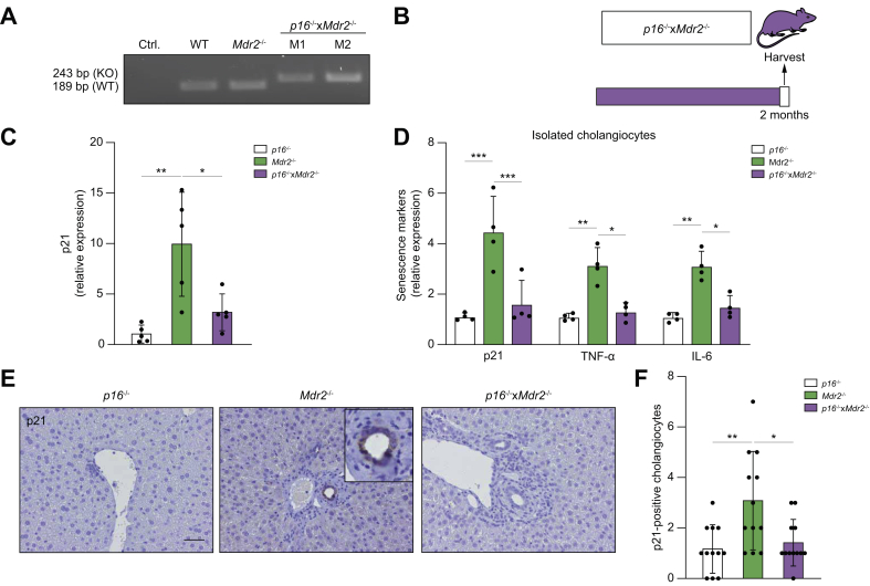
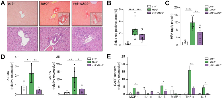
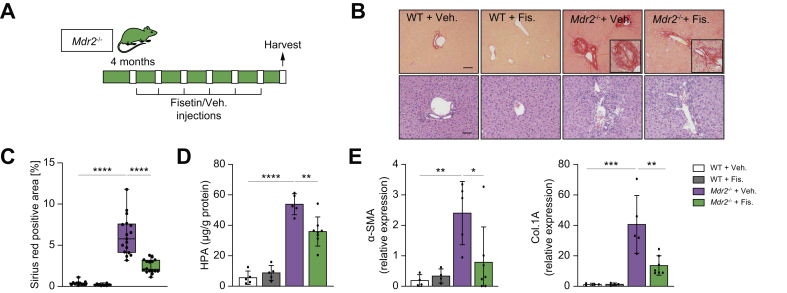
We used 2 novel genetic murine models to reduce cholangiocyte senescence: (i) p16 apoptosis through targeted activation of caspase (INK-ATTAC)x , in which the dimerizing molecule AP20187 promotes selective apoptotic removal of -expressing cells; and (ii) mice deficient in both and . mice were also treated with fisetin, a flavonoid molecule that selectively kills senescent cells. p16, p21, and inflammatory markers (tumour necrosis factor [TNF]-α, IL-1β, and monocyte chemoattractant protein-1 [MCP-1]) were measured by PCR, and hepatic fibrosis via a hydroxyproline assay and Sirius red staining.
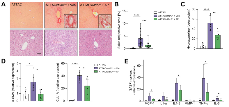
AP20187 treatment reduced p16 and p21 expression by 35% and ~70% ( >0.05), respectively. Expression of inflammatory markers (TNF-α, IL-1β, and MCP-1) decreased (by 60%, 40%, and 60%, respectively), and fibrosis was reduced by ~60% ( >0.05). Similarly, mice exhibited reduced p21 expression (70%), decreased expression of TNF-α, IL-1β (60%), and MCP-1 (65%) and reduced fibrosis (50%) ( >0.05) compared with mice. Fisetin treatment reduced expression of p16 and p21 (80% and 90%, respectively), TNF-α (50%), IL-1β (50%), MCP-1 (70%), and fibrosis (60%) ( >0.05).
Our data support a pathophysiological role of cholangiocyte senescence in the progression of PSC, and that targeted removal of senescent cholangiocytes is a plausible therapeutic approach.
Primary sclerosing cholangitis is a fibroinflammatory, incurable biliary disease. We previously reported that biliary epithelial cell senescence (cell-cycle arrest and hypersecretion of profibrotic molecules) is an important phenotype in primary sclerosing cholangitis. Herein, we demonstrate that reducing the number of senescent cholangiocytes leads to a reduction in the expression of inflammatory, fibrotic, and senescence markers associated with the disease.
胆管细胞衰老在原发性硬化性胆管炎(PSC)的发病机制中起重要作用。我们发现,细胞周期蛋白依赖性激酶抑制剂及衰老调节因子CDKN2A(p16)在PSC患者及PSC小鼠模型(多药耐药蛋白2缺陷小鼠)的胆管细胞中表达增加。鉴于近期数据表明减少衰老细胞对不同疾病有益,我们推测抑制胆管细胞衰老可改善多药耐药蛋白2缺陷小鼠的疾病状况。
我们使用2种新型基因小鼠模型来减少胆管细胞衰老:(i)通过靶向激活半胱天冬酶使p16凋亡(INK-ATTAC),其中二聚化分子AP20187可促进选择性凋亡清除表达p16的细胞;(ii)同时缺失多药耐药蛋白2和p16的小鼠。多药耐药蛋白2缺陷小鼠也用漆黄素进行处理,漆黄素是一种可选择性杀死衰老细胞的类黄酮分子。通过聚合酶链反应检测p16、p21及炎症标志物(肿瘤坏死因子[TNF]-α、白细胞介素-1β和单核细胞趋化蛋白-1[MCP-1]),并通过羟脯氨酸测定和天狼星红染色检测肝纤维化。
AP20187处理分别使p16和p21表达降低约35%和约70%(P>0.05)。炎症标志物(TNF-α、白细胞介素-1β和MCP-1)的表达下降(分别下降60%、40%和60%),纤维化降低约60%(P>0.05)。同样,与多药耐药蛋白2缺陷小鼠相比,同时缺失多药耐药蛋白2和p16的小鼠p21表达降低(70%),TNF-α、白细胞介素-1β(60%)和MCP-1(65%)的表达下降,纤维化降低(约50%)(P>0.05)。漆黄素处理使p16和p21表达降低(分别为80%和90%),TNF-α(50%)、白细胞介素-1β(50%)、MCP-1(70%)及纤维化(60%)降低(P>0.05)。
我们的数据支持胆管细胞衰老在PSC进展中的病理生理作用,且靶向清除衰老胆管细胞是一种可行的治疗方法。
原发性硬化性胆管炎是一种纤维炎症性、无法治愈的胆道疾病。我们之前报道,胆管上皮细胞衰老(细胞周期停滞和促纤维化分子的高分泌)是原发性硬化性胆管炎的一种重要表型。在此,我们证明减少衰老胆管细胞的数量可导致与该疾病相关的炎症、纤维化和衰老标志物的表达降低。