Liu Wei, Zhang Yifei, Nie Yechen, Liu Yifu, Li Zhongqi, Zhang Zhicheng, Gong Binbin, Ma Ming
Department of Urology, Gaoxin Branch of The First Affiliated Hospital of Nanchang University, Nanchang, 330000, China.
Jiangxi Provincial Key Laboratory of Urinary System Diseases, Department of Urology, The First Affiliated Hospital, Jiangxi Medical College, Nanchang University, Nanchang, Jiangxi, China.
Heliyon. 2024 Sep 4;10(18):e37086. doi: 10.1016/j.heliyon.2024.e37086. eCollection 2024 Sep 30.
AGBL2's role in tumorigenesis and cancer progression has been reported in several cancer studies, and it is closely associated with α-tubulin detyrosination. The roles of AGBL2 and α-tubulin detyrosination in renal cell carcinoma (RCC) pathogenesis remain unclear and require further investigation.
In this study, we conducted an analysis of AGBL2 expression differences between renal clear cell carcinoma tissues and normal tissues using data from The Cancer Genome Atlas (TCGA). We performed a comprehensive prognostic analysis of AGBL2 in Kidney Renal Clear Cell Carcinoma (KIRC) using univariate and multivariate Cox regression. Based on the results of the Cox analysis, we constructed a prognostic model to assess its predictive capabilities. Receiver Operating Characteristic (ROC) analysis confirmed the diagnostic value of AGBL2 in renal cancer. We conducted further validation by analyzing cancer tissue samples and renal cancer cell lines, which confirmed the role of AGBL2 in promoting RCC cell proliferation and migration through in vitro experiments. Additionally, we verified the impact of AGBL2's detyrosination on α-tubulin using the tubulin carboxypeptidase (TCP) inhibitor parthenolide. Finally, we performed sequencing analysis on AGBL2 knockdown 786-O cells to investigate the correlation between AGBL2, immune infiltration, and AKT phosphorylation. Moreover, we experimentally demonstrated the enhancing effect of AGBL2 on AKT phosphorylation.
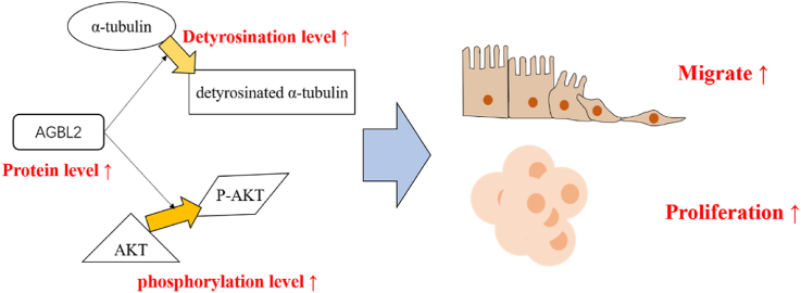
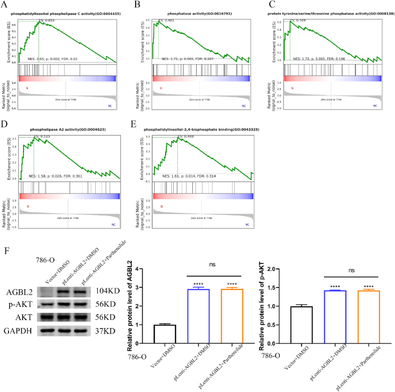
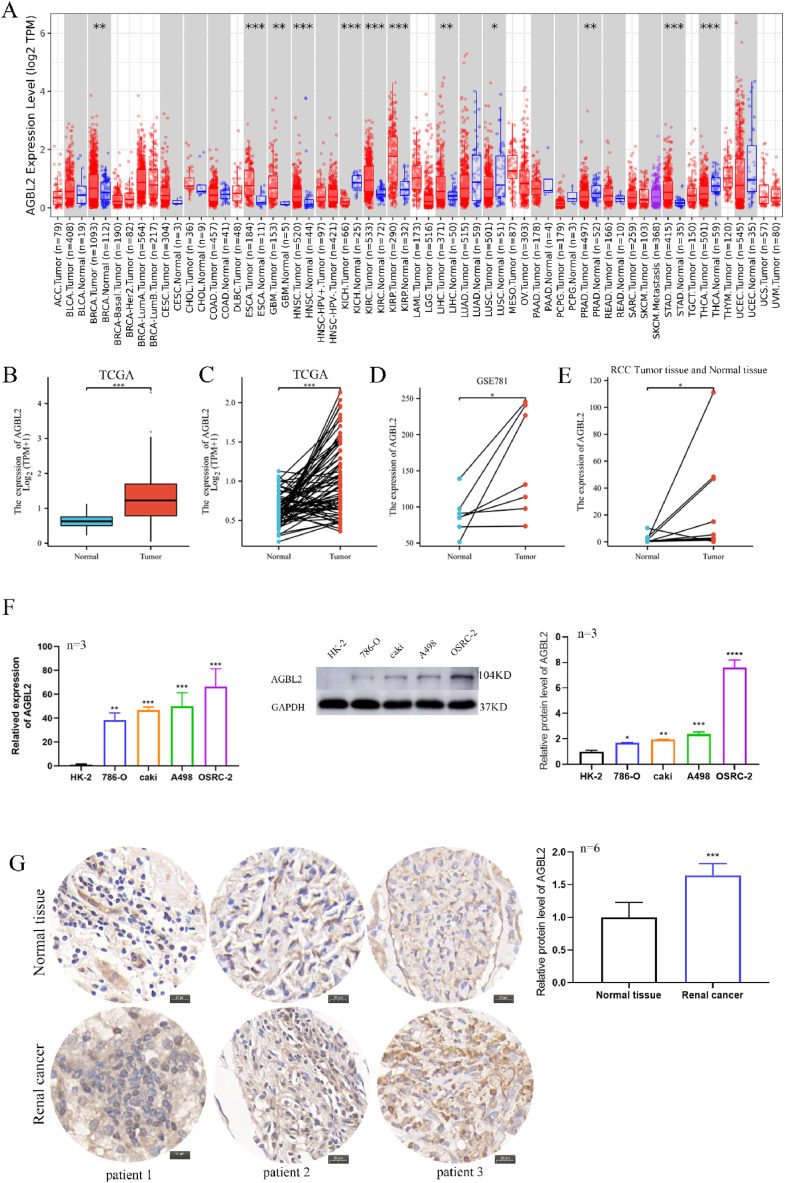
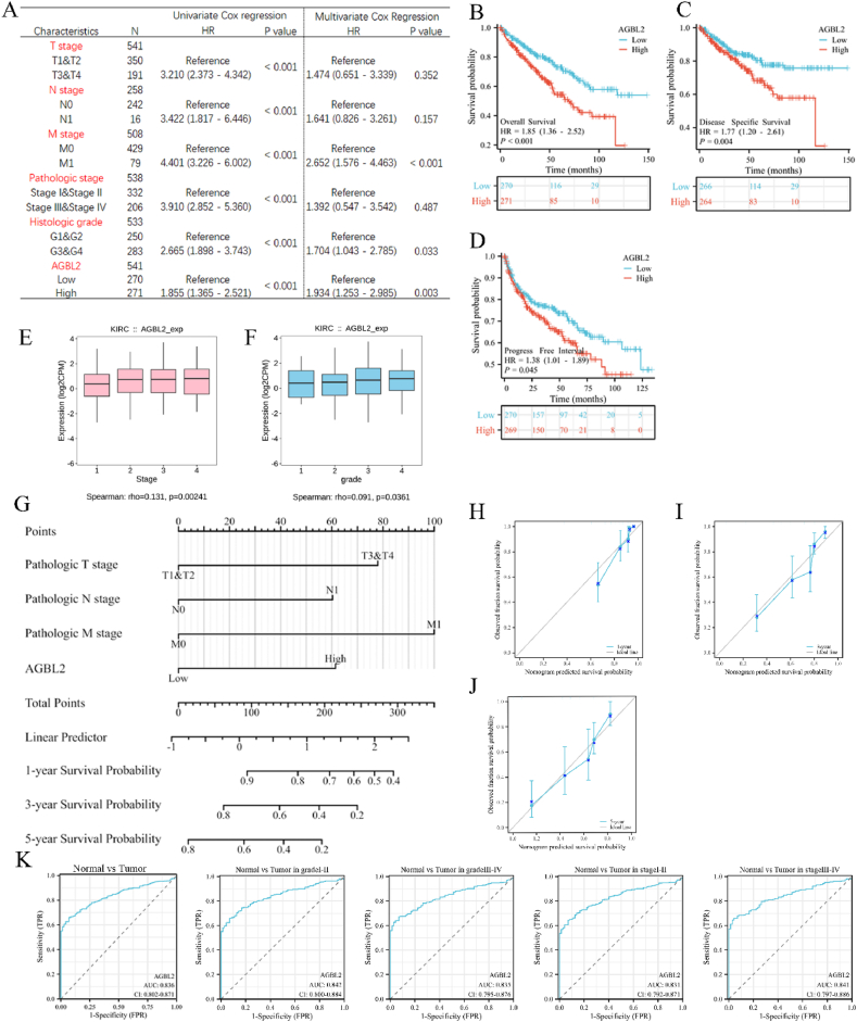
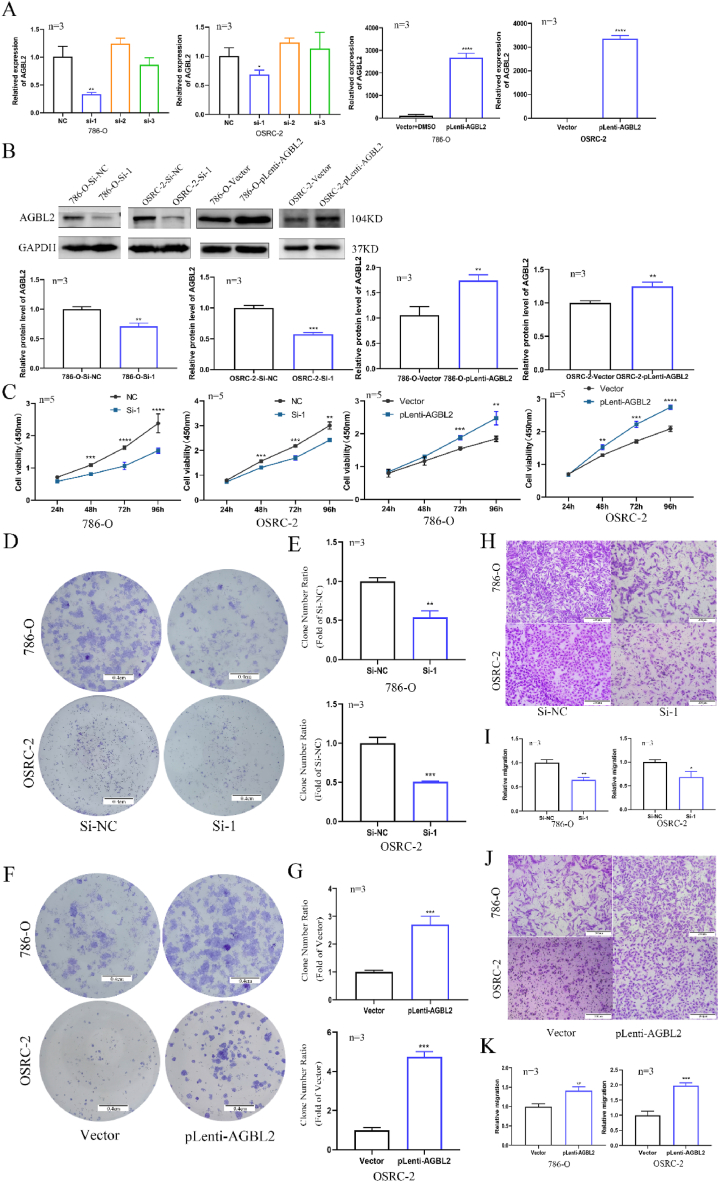
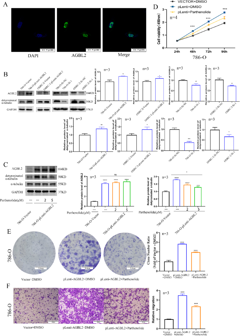
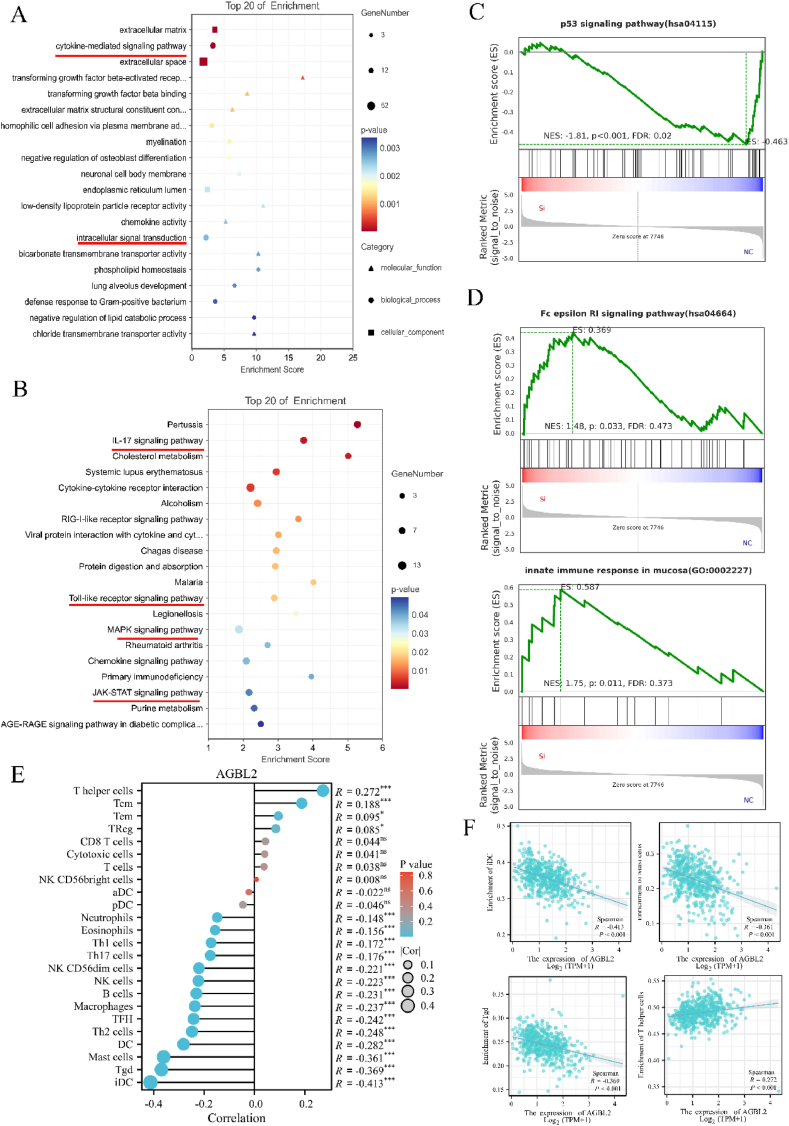
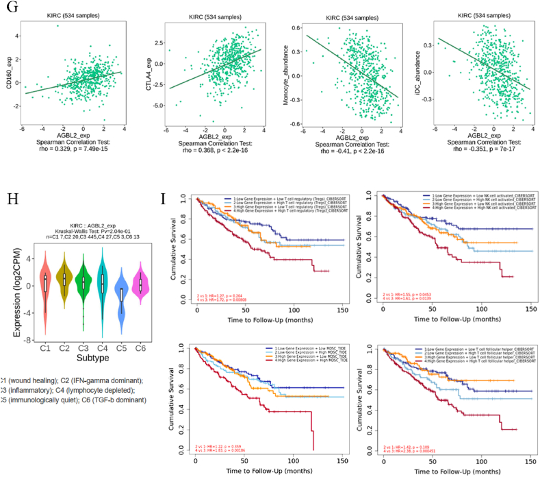
TCGA analysis revealed a significant increase in AGBL2 expression in RCC patients, which was correlated with poorer overall survival (OS), disease-specific survival (DSS), and progression-free intervals (PFI). According to the analysis results, we constructed column-line plots to predict the 1-, 3-, and 5-year survival outcomes in RCC patients. Additionally, the calibration plots assessing the model's performance exhibited favorable agreement with the predicted outcomes. And the ROC curves showed that AGBL2 showed good diagnostic performance in KIRC (AUC = 0.836)). Cell phenotyping assays revealed that AGBL2 knockdown in RCC cells significantly inhibited cell proliferation and migration. Conversely, overexpression of AGBL2 resulted in increased cell proliferation and migration in RCC cells. We observed that AGBL2 is predominantly located in the nucleus and can elevate the detyrosination level of α-tubulin in RCC cells. Moreover, the enhancement of RCC cell proliferation and migration by AGBL2 was partially inhibited after treatment with the TCP inhibitor parthenolide. Analysis of the sequencing data revealed that AGBL2 is associated with a diverse array of biological processes, encompassing signal transduction and immune infiltration. Interestingly, AGBL2 expression exhibited a negative correlation with the majority of immune cell infiltrations. Additionally, AGBL2 was found to enhance the phosphorylation of AKT in RCC cells.
Our study suggests that AGBL2 fosters RCC cell proliferation and migration by enhancing α-tubulin detyrosination. Moreover, elevated AGBL2 expression increases phosphorylation of AKT in RCC cells.
在多项癌症研究中已报道AGBL2在肿瘤发生和癌症进展中的作用,且它与α-微管蛋白去酪氨酸化密切相关。AGBL2和α-微管蛋白去酪氨酸化在肾细胞癌(RCC)发病机制中的作用仍不清楚,需要进一步研究。
在本研究中,我们使用来自癌症基因组图谱(TCGA)的数据,分析肾透明细胞癌组织和正常组织之间AGBL2表达差异。我们使用单变量和多变量Cox回归对肾透明细胞癌(KIRC)中的AGBL2进行全面的预后分析。基于Cox分析结果,我们构建了一个预后模型以评估其预测能力。受试者工作特征(ROC)分析证实了AGBL2在肾癌中的诊断价值。我们通过分析癌组织样本和肾癌细胞系进行进一步验证,通过体外实验证实了AGBL2在促进RCC细胞增殖和迁移中的作用。此外,我们使用微管蛋白羧肽酶(TCP)抑制剂小白菊内酯验证了AGBL2去酪氨酸化对α-微管蛋白的影响。最后,我们对AGBL2敲低的786-O细胞进行测序分析,以研究AGBL2、免疫浸润和AKT磷酸化之间的相关性。此外,我们通过实验证明了AGBL2对AKT磷酸化的增强作用。
TCGA分析显示RCC患者中AGBL2表达显著增加,这与较差的总生存期(OS)、疾病特异性生存期(DSS)和无进展生存期(PFI)相关。根据分析结果,我们构建了柱状线图以预测RCC患者1年、3年和5年的生存结果。此外,评估模型性能的校准图与预测结果显示出良好的一致性。并且ROC曲线表明AGBL2在KIRC中表现出良好的诊断性能(AUC = 0.836)。细胞表型分析显示,RCC细胞中AGBL2敲低显著抑制细胞增殖和迁移。相反,AGBL2的过表达导致RCC细胞中细胞增殖和迁移增加。我们观察到AGBL2主要位于细胞核中,并可提高RCC细胞中α-微管蛋白的去酪氨酸化水平。此外,用TCP抑制剂小白菊内酯处理后,AGBL2对RCC细胞增殖和迁移的增强作用部分受到抑制。对测序数据的分析表明,AGBL2与多种生物过程相关,包括信号转导和免疫浸润。有趣的是,AGBL2表达与大多数免疫细胞浸润呈负相关。此外,发现AGBL2可增强RCC细胞中AKT的磷酸化。
我们的研究表明,AGBL2通过增强α-微管蛋白去酪氨酸化促进RCC细胞增殖和迁移。此外,AGBL2表达升高会增加RCC细胞中AKT的磷酸化。