• 文献检索
  • 文档翻译
  • 深度研究
  • 学术资讯
  • Suppr Zotero 插件Zotero 插件
  • 邀请有礼
  • 套餐&价格
  • 历史记录
应用&插件
Suppr Zotero 插件Zotero 插件浏览器插件Mac 客户端Windows 客户端微信小程序
定价
高级版会员购买积分包购买API积分包
服务
文献检索文档翻译深度研究API 文档MCP 服务
关于我们
关于 Suppr公司介绍联系我们用户协议隐私条款
关注我们

Suppr 超能文献

核心技术专利:CN118964589B侵权必究
粤ICP备2023148730 号-1Suppr @ 2026

文献检索

告别复杂PubMed语法,用中文像聊天一样搜索,搜遍4000万医学文献。AI智能推荐,让科研检索更轻松。

立即免费搜索

文件翻译

保留排版,准确专业,支持PDF/Word/PPT等文件格式,支持 12+语言互译。

免费翻译文档

深度研究

AI帮你快速写综述,25分钟生成高质量综述,智能提取关键信息,辅助科研写作。

立即免费体验

一种基于超声检查和临床特征的实用风险分层系统,用于预测软组织肿块的恶性程度。

A practical risk stratification system based on ultrasonography and clinical characteristics for predicting the malignancy of soft tissue masses.

作者信息

Zhang Ying-Lun, Wu Meng-Jie, Hu Yu, Peng Xiao-Jing, Ma Qian, Mao Cui-Lian, Dong Ye, Wei Zong-Kai, Gao Ying-Qian, Yao Qi-Yu, Yao Jing, Ye Xin-Hua, Li Ju-Ming, Li Ao

机构信息

Department of Ultrasound, The First Affiliated Hospital of Nanjing Medical University, Nanjing, China.

Department of Ultrasound, The Affiliated Drum Tower Hospital of Nanjing University Medical School, Nanjing, China.

出版信息

Insights Imaging. 2024 Sep 19;15(1):226. doi: 10.1186/s13244-024-01802-9.

DOI:10.1186/s13244-024-01802-9
PMID:39320574
原文链接:https://pmc.ncbi.nlm.nih.gov/articles/PMC11424597/
Abstract

OBJECTIVE

To establish a practical risk stratification system (RSS) based on ultrasonography (US) and clinical characteristics for predicting soft tissue masses (STMs) malignancy.

METHODS

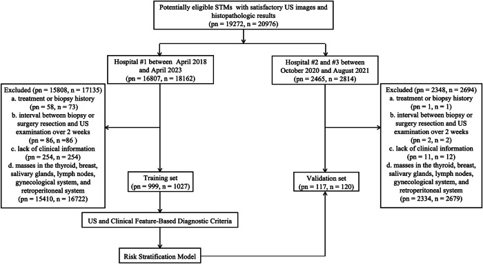
This retrospective multicenter study included patients with STMs who underwent US and pathological examinations between April 2018 and April 2023. Chi-square tests and multivariable logistic regression analyses were performed to assess the association of US and clinical characteristics with the malignancy of STMs in the training set. The RSS was constructed based on the scores of risk factors and validated externally.

RESULTS

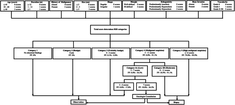
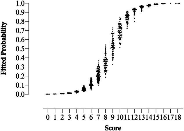
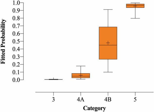
The training and validation sets included 1027 STMs (mean age, 50.90 ± 16.64, 442 benign and 585 malignant) and 120 STMs (mean age, 51.93 ± 17.90, 69 benign and 51 malignant), respectively. The RSS was constructed based on three clinical characteristics (age, duration, and history of malignancy) and six US characteristics (size, shape, margin, echogenicity, bone invasion, and vascularity). STMs were assigned to six categories in the RSS, including no abnormal findings, benign, probably benign (fitted probabilities [FP] for malignancy: 0.001-0.008), low suspicion (FP: 0.008-0.365), moderate suspicion (FP: 0.189-0.911), and high suspicion (FP: 0.798-0.999) for malignancy. The RSS displayed good diagnostic performance in the training and validation sets with area under the receiver operating characteristic curve (AUC) values of 0.883 and 0.849, respectively.

CONCLUSION

The practical RSS based on US and clinical characteristics could be useful for predicting STM malignancy, thereby providing the benefit of timely treatment strategy management to STM patients.

CRITICAL RELEVANCE STATEMENT

With the help of the RSS, better communication between radiologists and clinicians can be realized, thus facilitating tumor management.

KEY POINTS

There is no recognized grading system for STM management. A stratification system based on US and clinical features was built. The system realized great communication between radiologists and clinicians in tumor management.

摘要

目的

建立一种基于超声(US)和临床特征的实用风险分层系统(RSS),用于预测软组织肿块(STM)的恶性程度。

方法

这项回顾性多中心研究纳入了2018年4月至2023年4月期间接受US检查和病理检查的STM患者。在训练集中进行卡方检验和多变量逻辑回归分析,以评估US和临床特征与STM恶性程度的关联。基于风险因素得分构建RSS并进行外部验证。

结果

训练集和验证集分别包括1027个STM(平均年龄50.90±16.64岁,442个良性和585个恶性)和120个STM(平均年龄51.93±17.90岁,69个良性和51个恶性)。RSS基于三个临床特征(年龄、病程和恶性肿瘤病史)和六个US特征(大小、形状、边界、回声性、骨侵犯和血管情况)构建。STM在RSS中被分为六类,包括无异常发现、良性、可能良性(恶性的拟合概率[FP]:0.001 - 0.008)、低怀疑(FP:0.008 - 0.365)、中度怀疑(FP:0.189 - 0.911)和高怀疑(FP:0.798 - 0.999)恶性。RSS在训练集和验证集中表现出良好的诊断性能,受试者操作特征曲线(AUC)下面积值分别为0.883和0.849。

结论

基于US和临床特征的实用RSS可用于预测STM恶性程度,从而为STM患者提供及时治疗策略管理的益处。

关键相关性声明

借助RSS,放射科医生和临床医生之间可以实现更好的沟通,从而促进肿瘤管理。

要点

目前尚无公认的STM管理分级系统。建立了基于US和临床特征的分层系统。该系统在肿瘤管理中实现了放射科医生和临床医生之间的良好沟通。

https://cdn.ncbi.nlm.nih.gov/pmc/blobs/2ac5/11424597/df1aaf1009c9/13244_2024_1802_Fig4_HTML.jpg
https://cdn.ncbi.nlm.nih.gov/pmc/blobs/2ac5/11424597/3b608034a568/13244_2024_1802_Fig1_HTML.jpg
https://cdn.ncbi.nlm.nih.gov/pmc/blobs/2ac5/11424597/6c12124f0063/13244_2024_1802_Fig2_HTML.jpg
https://cdn.ncbi.nlm.nih.gov/pmc/blobs/2ac5/11424597/1de11e35eb6c/13244_2024_1802_Fig3_HTML.jpg
https://cdn.ncbi.nlm.nih.gov/pmc/blobs/2ac5/11424597/df1aaf1009c9/13244_2024_1802_Fig4_HTML.jpg

相似文献

1
A practical risk stratification system based on ultrasonography and clinical characteristics for predicting the malignancy of soft tissue masses.一种基于超声检查和临床特征的实用风险分层系统,用于预测软组织肿块的恶性程度。
Insights Imaging. 2024 Sep 19;15(1):226. doi: 10.1186/s13244-024-01802-9.
2
Nomogram Based on Ultrasonography and Clinical Features for Predicting Malignancy in Soft Tissue Tumors.基于超声检查和临床特征的软组织肿瘤恶性风险预测列线图
Cancer Manag Res. 2021 Mar 2;13:2143-2152. doi: 10.2147/CMAR.S296972. eCollection 2021.
3
Diagnostic Performance of US and MRI in Predicting Malignancy of Soft Tissue Masses: Using a Scoring System.超声和磁共振成像在预测软组织肿块恶性程度中的诊断性能:采用评分系统
Front Oncol. 2022 Apr 29;12:853232. doi: 10.3389/fonc.2022.853232. eCollection 2022.
4
Validation of Ultrasound Risk Stratification Systems for Cervical Lymph Node Metastasis in Patients with Thyroid Cancer.甲状腺癌患者颈部淋巴结转移超声风险分层系统的验证
Cancers (Basel). 2022 Apr 23;14(9):2106. doi: 10.3390/cancers14092106.
5
Predicting the risk of malignancy in adnexal masses based on the Simple Rules from the International Ovarian Tumor Analysis group.基于国际卵巢肿瘤分析小组的简单规则预测附件包块的恶性风险。
Am J Obstet Gynecol. 2016 Apr;214(4):424-437. doi: 10.1016/j.ajog.2016.01.007. Epub 2016 Jan 19.
6
Soft tissue masses: distribution of entities and rate of malignancy in small lesions.软组织肿块:小病变实体的分布和恶性率。
BMC Cancer. 2021 Jan 22;21(1):93. doi: 10.1186/s12885-020-07769-2.
7
Web-Based Malignancy Risk Estimation for Thyroid Nodules Using Ultrasonography Characteristics: Development and Validation of a Predictive Model.基于超声特征的甲状腺结节网络恶性风险评估:预测模型的开发与验证
Thyroid. 2015 Dec;25(12):1306-12. doi: 10.1089/thy.2015.0188. Epub 2015 Oct 26.
8
The diagnostic accuracy of intravoxel incoherent motion and diffusion kurtosis imaging in the differentiation of malignant and benign soft-tissue masses: which is better?磁共振体素内不相干运动和扩散峰度成像诊断良恶性软组织肿块的准确性:哪个更好?
Acta Radiol. 2022 Jun;63(6):785-793. doi: 10.1177/02841851211017511. Epub 2021 May 17.
9
Does size reliably predict malignancy in soft tissue tumours?软组织肿瘤的大小能可靠地预测恶性肿瘤吗?
Eur Radiol. 2016 Dec;26(12):4640-4648. doi: 10.1007/s00330-016-4300-z. Epub 2016 Mar 9.
10
Contrast-enhanced US to Improve Diagnostic Performance of O-RADS US Risk Stratification System for Malignancy.超声造影增强有助于提高 O-RADS-US 风险分层系统对恶性肿瘤的诊断性能。
Radiology. 2023 Aug;308(2):e223003. doi: 10.1148/radiol.223003.

本文引用的文献

1
Benefit of Using Both Ultrasound Imaging and Clinical Information for Predicting Malignant Soft Tissue Tumors.超声成像与临床信息联合应用预测软组织恶性肿瘤的获益。
Ultrasound Med Biol. 2023 Dec;49(12):2459-2468. doi: 10.1016/j.ultrasmedbio.2023.08.005. Epub 2023 Sep 11.
2
A multiparametric clinic-ultrasomics nomogram for predicting extremity soft-tissue tumor malignancy: a combined retrospective and prospective bicentric study.多参数临床-超声组学列线图预测肢体软组织肿瘤恶性程度:一项回顾性和前瞻性的中心联合研究。
Radiol Med. 2023 Jun;128(6):784-797. doi: 10.1007/s11547-023-01639-0. Epub 2023 May 8.
3
Musculoskeletal Soft-tissue Masses: MR imaging-Ultrasonography Correlation, with an Emphasis on the 2020 World Health Organization Classification.
肌肉骨骼软组织肿块:磁共振成像-超声影像学相关性,重点介绍 2020 年世界卫生组织分类。
Magn Reson Imaging Clin N Am. 2023 May;31(2):285-308. doi: 10.1016/j.mric.2022.10.001. Epub 2023 Feb 4.
4
Does the apparent diffusion coefficient from diffusion-weighted MRI imaging aid in the characterization of malignant soft tissue tumors and sarcomas.弥散加权 MRI 成像的表观扩散系数是否有助于恶性软组织肿瘤和肉瘤的特征化。
Skeletal Radiol. 2023 Aug;52(8):1475-1484. doi: 10.1007/s00256-023-04289-5. Epub 2023 Feb 2.
5
Added value of contrast-enhanced ultrasound to conventional ultrasound for characterization of indeterminate soft-tissue tumors.对比增强超声在常规超声对不确定软组织肿瘤的特征化中的应用价值。
Br J Radiol. 2023 Jan 1;96(1141):20220404. doi: 10.1259/bjr.20220404. Epub 2022 Dec 9.
6
A Practical CEUS Thyroid Reporting System for Thyroid Nodules.甲状腺结节超声造影实用报告系统
Radiology. 2022 Oct;305(1):149-159. doi: 10.1148/radiol.212319. Epub 2022 Jun 14.
7
Diagnostic Performance of US and MRI in Predicting Malignancy of Soft Tissue Masses: Using a Scoring System.超声和磁共振成像在预测软组织肿块恶性程度中的诊断性能:采用评分系统
Front Oncol. 2022 Apr 29;12:853232. doi: 10.3389/fonc.2022.853232. eCollection 2022.
8
Management of Soft-tissue Tumors With a Size of 2-5 cm, Including Malignancy.2-5cm 大小软组织肿瘤(包含恶性肿瘤)的处理。
Anticancer Res. 2022 Mar;42(3):1555-1562. doi: 10.21873/anticanres.15629.
9
Qualitative and Quantitative Contrast-Enhanced Ultrasound Combined with Conventional Ultrasound for Predicting the Malignancy of Soft Tissue Tumors.定性和定量对比增强超声联合常规超声预测软组织肿瘤的恶性程度。
Ultrasound Med Biol. 2022 Feb;48(2):237-247. doi: 10.1016/j.ultrasmedbio.2021.10.007. Epub 2021 Nov 12.
10
Nomogram Based on Ultrasonography and Clinical Features for Predicting Malignancy in Soft Tissue Tumors.基于超声检查和临床特征的软组织肿瘤恶性风险预测列线图
Cancer Manag Res. 2021 Mar 2;13:2143-2152. doi: 10.2147/CMAR.S296972. eCollection 2021.