Zhang Lindy, Maalouf Alexandre, Makri Stavriani C, Banerjee Jineta, Suru Aditya, Tam Ada J, Calizo Ana, Pollard Kai, Wang Jiawan, Danilova Ludmila, Ioannou Maria, Levin Adam S, Morris Carol D, Rhee Daniel S, Belzberg Allan J, Blakeley Jaishri O, Ladle Brian H, Pardoll Drew M, Lucas Calixto-Hope G, Rodriguez Fausto J, Gross John M, Anders Robert A, Pratilas Christine A, Llosa Nicolas J
Department of Oncology, Sidney Kimmel Comprehensive Cancer Center, Johns Hopkins University School of Medicine, Baltimore, Maryland.
Cellular and Molecular Medicine Graduate Program, Johns Hopkins University School of Medicine, Baltimore, Maryland.
Clin Cancer Res. 2024 Dec 2;30(23):5459-5472. doi: 10.1158/1078-0432.CCR-24-1454.
Malignant peripheral nerve sheath tumors (MPNST) are aggressive soft-tissue sarcomas and the leading cause of mortality in individuals with neurofibromatosis type 1 (NF1). Despite many clinical trials, outcomes for patients with MPNST have remained stagnant, and most succumb to their disease; thus, novel therapeutic approaches are needed. A better understanding of the MPNST immune ecosystem will aid in the development of strategies to activate the immune system against the tumor. In this study, we profile the tumor immune microenvironment (TIME) in NF1-associated peripheral nerve sheath tumors (PNST) to discover insights on the role played by tumor-infiltrating immune cells in malignant transformation.
Using fresh and formalin-fixed paraffin-embedded tissue from patients diagnosed with NF1-PNST, we dissected the TIME through IHC, multiparameter flow cytometry, and comparative transcriptomic studies.
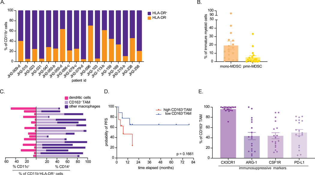
Immunophenotyping confirmed increased immune cell infiltration during malignant progression, with a predominance of infiltrating myeloid cells, particularly CD163+ tumor-associated macrophages (TAM). The T cells within MPNST exhibited signs of tumor activation, characterized by high programmed cell death 1 expression. Additionally, MPNST specimens demonstrated elevated levels of immunosuppressive TAM, with heightened PD-L1 expression. The proportion of CD163+ myeloid cells within the TIME correlated with poorer progression-free survival. Notably, loss of H3K27 trimethylation correlated with low immune cell infiltration in MPNST.
Malignant transformation of NF1-PNST is characterized by an immunosuppressive microenvironment comprising TAM with high expression of PD-L1, which is associated with inferior outcomes. These findings suggest the clinical potential of immune-modulating therapeutics that can unleash an antitumor immune response.
恶性外周神经鞘瘤(MPNST)是侵袭性软组织肉瘤,也是1型神经纤维瘤病(NF1)患者死亡的主要原因。尽管进行了许多临床试验,但MPNST患者的治疗结果一直停滞不前,大多数患者最终死于该疾病;因此,需要新的治疗方法。更好地了解MPNST免疫生态系统将有助于制定激活免疫系统对抗肿瘤的策略。在本研究中,我们分析了NF1相关外周神经鞘瘤(PNST)的肿瘤免疫微环境(TIME),以了解肿瘤浸润免疫细胞在恶性转化中所起的作用。
我们使用来自诊断为NF1-PNST患者的新鲜和福尔马林固定石蜡包埋组织,通过免疫组化、多参数流式细胞术和比较转录组学研究剖析TIME。
免疫表型分析证实,在恶性进展过程中免疫细胞浸润增加,主要是浸润的髓系细胞,尤其是CD163+肿瘤相关巨噬细胞(TAM)。MPNST内的T细胞表现出肿瘤激活的迹象,其特征是程序性细胞死亡1表达水平高。此外,MPNST标本显示免疫抑制性TAM水平升高,PD-L1表达增强。TIME内CD163+髓系细胞的比例与无进展生存期较差相关。值得注意的是,H3K27三甲基化的缺失与MPNST中免疫细胞浸润低相关。
NF1-PNST的恶性转化特征是免疫抑制微环境,包括高表达PD-L1的TAM,这与较差的治疗结果相关。这些发现提示了能够引发抗肿瘤免疫反应的免疫调节疗法的临床潜力。