• 文献检索
  • 文档翻译
  • 深度研究
  • 学术资讯
  • Suppr Zotero 插件Zotero 插件
  • 邀请有礼
  • 套餐&价格
  • 历史记录
应用&插件
Suppr Zotero 插件Zotero 插件浏览器插件Mac 客户端Windows 客户端微信小程序
定价
高级版会员购买积分包购买API积分包
服务
文献检索文档翻译深度研究API 文档MCP 服务
关于我们
关于 Suppr公司介绍联系我们用户协议隐私条款
关注我们

Suppr 超能文献

核心技术专利:CN118964589B侵权必究
粤ICP备2023148730 号-1Suppr @ 2026

文献检索

告别复杂PubMed语法,用中文像聊天一样搜索,搜遍4000万医学文献。AI智能推荐,让科研检索更轻松。

立即免费搜索

文件翻译

保留排版,准确专业,支持PDF/Word/PPT等文件格式,支持 12+语言互译。

免费翻译文档

深度研究

AI帮你快速写综述,25分钟生成高质量综述,智能提取关键信息,辅助科研写作。

立即免费体验

牙硬组织应力分布的有限元方法研究:洞型设计与修复材料的影响

A Finite Element Method Study of Stress Distribution in Dental Hard Tissues: Impact of Access Cavity Design and Restoration Material.

作者信息

Boțilă Mihaela-Roxana, Popa Dragos Laurențiu, Mercuț Răzvan, Iacov-Crăițoiu Monica Mihaela, Scrieciu Monica, Popescu Sanda Mihaela, Mercuț Veronica

机构信息

Department of Prosthetic Dentistry, University of Medicine and Pharmacy of Craiova, 200349 Craiova, Romania.

Department of Automotive, Transportation and Industrial Engineering, Faculty of Mechanics, University of Craiova, 200478 Craiova, Romania.

出版信息

Bioengineering (Basel). 2024 Aug 29;11(9):878. doi: 10.3390/bioengineering11090878.

DOI:10.3390/bioengineering11090878
PMID:39329620
原文链接:https://pmc.ncbi.nlm.nih.gov/articles/PMC11429090/
Abstract

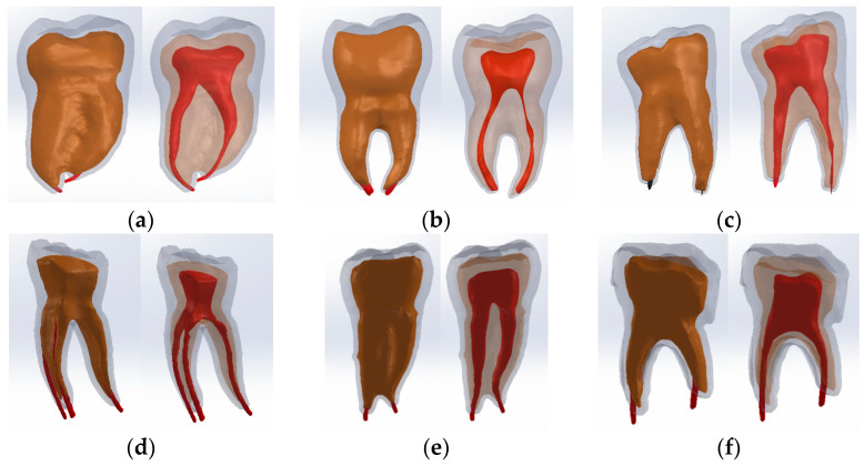
The design of the access cavity is an important factor in endodontic treatment for the further evolution of the tooth. The objective of this study was to highlight the most favorable access cavity design (TrussAC, UltraAC, TradAC, CariesAC, ConsAC, RestoAC) based on the stress distribution on virtual models of mandibular molars. To achieve the objectives of the study, four series of virtual models of six molars were made. The first two series of external virtual models were obtained based on the three-dimensional scanning of the molars before the access cavity preparation and after their restoration, to obtain the density of the restorative materials. Internal morphology was added to the next two series of virtual models and after that, materials were added, specific for root canal obturation and coronal restoration. The simulations were performed for two coronary restoration materials, bulk fill composite and amalgam. The results showed, based on the stress maps, that the highest values were recorded for CariesAC and the lowest values for UltraAC. Comparing the two restorative materials, the lowest level of stress, strains, and displacements was highlighted in the case of UltraAC, TradAC, and ConsAC cavities for amalgam. The results obtained in this study should guide doctors towards a conservative attitude with the preservation of as much hard tissue as possible and the differentiated use of restorative materials according to the amount of tissue lost when preparing the access cavity.

摘要

进入髓腔的设计是牙髓治疗中影响牙齿进一步发展的重要因素。本研究的目的是根据下颌磨牙虚拟模型上的应力分布,突出最有利的进入髓腔设计(桁架式髓腔、超常规髓腔、传统髓腔、龋坏性髓腔、保守性髓腔、修复性髓腔)。为实现本研究的目的,制作了四组共六个磨牙的虚拟模型。前两组外部虚拟模型是通过对磨牙在制备进入髓腔前和修复后的三维扫描获得的,以得到修复材料的密度。接下来的两组虚拟模型添加了内部形态,之后添加了用于根管充填和冠部修复的特定材料。针对两种冠部修复材料(大块充填复合树脂和汞合金)进行了模拟。基于应力图的结果显示,龋坏性髓腔记录到的应力值最高,而超常规髓腔的应力值最低。比较这两种修复材料,在使用汞合金时,超常规髓腔、传统髓腔和保守性髓腔的应力、应变和位移水平最低。本研究所得结果应引导医生采取保守态度,尽可能保留更多的硬组织,并根据制备进入髓腔时丢失的组织量区别使用修复材料。

https://cdn.ncbi.nlm.nih.gov/pmc/blobs/7d6c/11429090/33df2a7e6612/bioengineering-11-00878-g021.jpg
https://cdn.ncbi.nlm.nih.gov/pmc/blobs/7d6c/11429090/35081d73d855/bioengineering-11-00878-g018.jpg
https://cdn.ncbi.nlm.nih.gov/pmc/blobs/7d6c/11429090/7e0642377867/bioengineering-11-00878-g001.jpg
https://cdn.ncbi.nlm.nih.gov/pmc/blobs/7d6c/11429090/ce1afe12cf3f/bioengineering-11-00878-g002.jpg
https://cdn.ncbi.nlm.nih.gov/pmc/blobs/7d6c/11429090/34be1b515e30/bioengineering-11-00878-g003.jpg
https://cdn.ncbi.nlm.nih.gov/pmc/blobs/7d6c/11429090/69930fb26828/bioengineering-11-00878-g004.jpg
https://cdn.ncbi.nlm.nih.gov/pmc/blobs/7d6c/11429090/47fa8b23891e/bioengineering-11-00878-g005.jpg
https://cdn.ncbi.nlm.nih.gov/pmc/blobs/7d6c/11429090/16925c77f792/bioengineering-11-00878-g006.jpg
https://cdn.ncbi.nlm.nih.gov/pmc/blobs/7d6c/11429090/19234f837ecc/bioengineering-11-00878-g007.jpg
https://cdn.ncbi.nlm.nih.gov/pmc/blobs/7d6c/11429090/5c9ed0ed53aa/bioengineering-11-00878-g008.jpg
https://cdn.ncbi.nlm.nih.gov/pmc/blobs/7d6c/11429090/51ca0da7979f/bioengineering-11-00878-g009.jpg
https://cdn.ncbi.nlm.nih.gov/pmc/blobs/7d6c/11429090/36b82b9addb5/bioengineering-11-00878-g010.jpg
https://cdn.ncbi.nlm.nih.gov/pmc/blobs/7d6c/11429090/671713ca2d41/bioengineering-11-00878-g011.jpg
https://cdn.ncbi.nlm.nih.gov/pmc/blobs/7d6c/11429090/30da93877a4a/bioengineering-11-00878-g012.jpg
https://cdn.ncbi.nlm.nih.gov/pmc/blobs/7d6c/11429090/be6bfb035106/bioengineering-11-00878-g013.jpg
https://cdn.ncbi.nlm.nih.gov/pmc/blobs/7d6c/11429090/a8712e2cdfc5/bioengineering-11-00878-g014.jpg
https://cdn.ncbi.nlm.nih.gov/pmc/blobs/7d6c/11429090/175868680732/bioengineering-11-00878-g015.jpg
https://cdn.ncbi.nlm.nih.gov/pmc/blobs/7d6c/11429090/bb5f4b361ce6/bioengineering-11-00878-g016.jpg
https://cdn.ncbi.nlm.nih.gov/pmc/blobs/7d6c/11429090/fb151713b6f9/bioengineering-11-00878-g017.jpg
https://cdn.ncbi.nlm.nih.gov/pmc/blobs/7d6c/11429090/bbf8846bc37e/bioengineering-11-00878-g019.jpg
https://cdn.ncbi.nlm.nih.gov/pmc/blobs/7d6c/11429090/556dee696ead/bioengineering-11-00878-g020.jpg
https://cdn.ncbi.nlm.nih.gov/pmc/blobs/7d6c/11429090/33df2a7e6612/bioengineering-11-00878-g021.jpg

相似文献

1
A Finite Element Method Study of Stress Distribution in Dental Hard Tissues: Impact of Access Cavity Design and Restoration Material.牙硬组织应力分布的有限元方法研究:洞型设计与修复材料的影响
Bioengineering (Basel). 2024 Aug 29;11(9):878. doi: 10.3390/bioengineering11090878.
2
Comparative Evaluation of Different Endodontic Access Cavity Designs with Different Burs-An Experimental Insight into the Aspect of Fracture Resistance.不同车针对不同根管入口洞形设计的比较评价——抗折性方面的实验洞察
J Pharm Bioallied Sci. 2024 Feb;16(Suppl 1):S821-S824. doi: 10.4103/jpbs.jpbs_1038_23. Epub 2023 Dec 20.
3
Influence of Cavity Designs on Fracture Resistance: Analysis of the Role of Different Access Techniques to the Endodontic Cavity in the Onset of Fractures: Narrative Review.不同根管入路方式对根管治疗后牙体抗折性的影响:一种叙述性综述。
ScientificWorldJournal. 2024 Jul 30;2024:1648011. doi: 10.1155/2024/1648011. eCollection 2024.
4
Influence of ultraconservative access cavities on instrumentation efficacy with XP-endo Shaper and Reciproc, filling ability and load capacity of mandibular molars subjected to thermomechanical cycling.超保守开髓对 XP-endo Shaper 和 Reciproc 器械效能、下颌磨牙热机械循环后充填能力和承载能力的影响。
Int Endod J. 2021 Aug;54(8):1383-1393. doi: 10.1111/iej.13525. Epub 2021 Apr 26.
5
Current strategies for conservative endodontic access cavity preparation techniques-systematic review, meta-analysis, and decision-making protocol.当前保守性牙髓腔入口制备技术的策略——系统综述、荟萃分析和决策制定方案。
Clin Oral Investig. 2021 Nov;25(11):6027-6044. doi: 10.1007/s00784-021-04080-7. Epub 2021 Oct 8.
6
Preserving dentine in minimally invasive access cavities does not strength fracture resistance of restored mandibular molars.微创备洞时保留牙本质不会增强下颌磨牙修复后的抗折能力。
Int Endod J. 2021 Jun;54(6):966-974. doi: 10.1111/iej.13487. Epub 2021 Feb 19.
7
A Comparison of Volume of Tissue Removed and Biomechanical Analysis of Different Access Cavity Designs in 2-rooted Mandibular First Molars: A Multisample 3-dimensional Finite Element Analysis.不同下颌第一磨牙双根管根管治疗术不同入路预备后组织去除量和生物力学的三维有限元分析。
J Endod. 2022 Mar;48(3):362-369. doi: 10.1016/j.joen.2021.12.007. Epub 2021 Dec 22.
8
Effect of fiber-reinforced direct restorative materials on the fracture resistance of endodontically treated mandibular molars restored with a conservative endodontic cavity design.纤维增强型直接修复材料对采用保守根管治疗窝洞设计修复的下颌磨牙抗折性能的影响。
Clin Oral Investig. 2024 May 16;28(6):316. doi: 10.1007/s00784-024-05720-4.
9
Effect of guided conservative endodontic access and different file kinematics on debris extrusion in mesial root of the mandibular molars: An study.引导式保守性牙髓治疗入路及不同锉运动学对下颌磨牙近中根碎屑挤出的影响:一项研究。
J Conserv Dent. 2022 Sep-Oct;25(5):547-554. doi: 10.4103/jcd.jcd_273_22. Epub 2022 Sep 12.
10
Effect of Different Endodontic Access Cavity Designs in Combination with WaveOne Gold and TruNatomy on the Fracture Resistance of Mandibular First Molars: A Nonlinear Finite Element Analysis.不同开髓洞形设计联合WaveOne Gold和TruNatomy对下颌第一磨牙抗折性的影响:非线性有限元分析
J Endod. 2023 May;49(5):559-566. doi: 10.1016/j.joen.2023.03.004. Epub 2023 Mar 15.

引用本文的文献

1
The Effect of Stress Distribution by Fiberglass Post System With Different Designs on Endodontically Treated Maxillary Central Incisors: A Three-Dimensional Finite Element Analysis.不同设计的玻璃纤维桩系统对根管治疗后的上颌中切牙应力分布的影响:三维有限元分析
Cureus. 2025 Mar 31;17(3):e81545. doi: 10.7759/cureus.81545. eCollection 2025 Mar.
2
Stress Evaluation in Endodontically Treated Virtual Teeth Restored with Composite Fillings and Cast or Fiberglass Posts-A Finite Element Analysis.用复合树脂充填材料及铸造桩或玻璃纤维桩修复的根管治疗虚拟牙齿的应力评估——有限元分析
Biomedicines. 2025 Apr 16;13(4):974. doi: 10.3390/biomedicines13040974.

本文引用的文献

1
Stress distribution pattern in two different no-post systems in endodontically treated maxillary central incisors: A three-dimensional finite element analysis.根管治疗后的上颌中切牙两种不同无桩系统中的应力分布模式:三维有限元分析
J Conserv Dent Endod. 2024 Jun;27(6):572-576. doi: 10.4103/JCDE.JCDE_135_24. Epub 2024 Jun 6.
2
Effect of fiber-reinforced direct restorative materials on the fracture resistance of endodontically treated mandibular molars restored with a conservative endodontic cavity design.纤维增强型直接修复材料对采用保守根管治疗窝洞设计修复的下颌磨牙抗折性能的影响。
Clin Oral Investig. 2024 May 16;28(6):316. doi: 10.1007/s00784-024-05720-4.
3
Impact of Access Cavity Design on Fracture Resistance of Endodontically Treated Maxillary First Premolar: In Vitro.
根管治疗上颌第一前磨牙的牙体预备对其抗折性能的影响:体外研究。
Braz Dent J. 2024 Mar 22;35:e245676. doi: 10.1590/0103-6440202405676. eCollection 2024.
4
A Retrospective Study on the Fracture Toughness of the Coronal Restorations in Endodontically Restored Teeth. An Original Research.牙髓治疗后牙齿冠部修复体断裂韧性的回顾性研究。一项原创研究。
J Pharm Bioallied Sci. 2023 Jul;15(Suppl 1):S132-S136. doi: 10.4103/jpbs.jpbs_436_22. Epub 2023 Jul 5.
5
Stereomicroscopic Aspects of Non-Carious Cervical Lesions.非龋性颈部病变的体视显微镜观察
Diagnostics (Basel). 2023 Aug 3;13(15):2590. doi: 10.3390/diagnostics13152590.
6
Decision Making in the Restoration of Endodontically Treated Teeth: Effect of Biomimetic Dentistry Training.牙髓治疗后牙齿修复中的决策制定:仿生牙科培训的影响。
Dent J (Basel). 2023 Jun 26;11(7):159. doi: 10.3390/dj11070159.
7
Five-Year Clinical Performance of Complex Class II Resin Composite and Amalgam Restorations-A Retrospective Study.复杂II类树脂复合材料和银汞合金修复体的五年临床性能——一项回顾性研究
Dent J (Basel). 2023 Mar 24;11(4):88. doi: 10.3390/dj11040088.
8
Teeth Restored with Bulk-Fill Composites and Conventional Resin Composites; Investigation of Stress Distribution and Fracture Lifespan on Enamel, Dentin, and Restorative Materials via Three-Dimensional Finite Element Analysis.采用大块充填复合树脂和传统树脂复合材料修复的牙齿;通过三维有限元分析对牙釉质、牙本质和修复材料的应力分布及断裂寿命进行研究。
Polymers (Basel). 2023 Mar 25;15(7):1637. doi: 10.3390/polym15071637.
9
Long-term tooth survival and success following primary root canal treatment: a 5- to 37-year retrospective observation.一次性根管治疗后长期牙齿存活和成功的回顾性观察:5 年至 37 年的随访。
Clin Oral Investig. 2023 Jun;27(6):3233-3244. doi: 10.1007/s00784-023-04938-y. Epub 2023 Mar 18.
10
Stress distribution of endodontically treated mandibular molars with varying amounts of tooth structure restored with direct composite resin with or without cuspal coverage: A 3D finite element analysis.使用或不使用牙尖覆盖的直接复合树脂修复不同牙体结构量的根管治疗下颌磨牙的应力分布:三维有限元分析
J Conserv Dent. 2023 Jan-Feb;26(1):20-25. doi: 10.4103/jcd.jcd_333_22. Epub 2022 Dec 8.