• 文献检索
  • 文档翻译
  • 深度研究
  • 学术资讯
  • Suppr Zotero 插件Zotero 插件
  • 邀请有礼
  • 套餐&价格
  • 历史记录
应用&插件
Suppr Zotero 插件Zotero 插件浏览器插件Mac 客户端Windows 客户端微信小程序
定价
高级版会员购买积分包购买API积分包
服务
文献检索文档翻译深度研究API 文档MCP 服务
关于我们
关于 Suppr公司介绍联系我们用户协议隐私条款
关注我们

Suppr 超能文献

核心技术专利:CN118964589B侵权必究
粤ICP备2023148730 号-1Suppr @ 2026

文献检索

告别复杂PubMed语法,用中文像聊天一样搜索,搜遍4000万医学文献。AI智能推荐,让科研检索更轻松。

立即免费搜索

文件翻译

保留排版,准确专业,支持PDF/Word/PPT等文件格式,支持 12+语言互译。

免费翻译文档

深度研究

AI帮你快速写综述,25分钟生成高质量综述,智能提取关键信息,辅助科研写作。

立即免费体验

LUSBI 方案(肺部超声/ BREST 评分/下腔静脉)-其在心源性和非心源性呼吸困难鉴别诊断中的作用。

The LUSBI Protocol (Lung Ultrasound/BREST Score/Inferior Vena Cava)-Its Role in a Differential Diagnostic Approach to Dyspnea of Cardiogenic and Non-Cardiogenic Origin.

机构信息

Emergency Medical Service of Primary Health Care Center in Banja Luka, 78000 Banja Luka, Bosnia and Herzegovina.

Medical Faculty, University of Banja Luka, 78000 Banja Luka, Bosnia and Herzegovina.

出版信息

Medicina (Kaunas). 2024 Sep 18;60(9):1521. doi: 10.3390/medicina60091521.

DOI:10.3390/medicina60091521
PMID:39336562
原文链接:https://pmc.ncbi.nlm.nih.gov/articles/PMC11433694/
Abstract

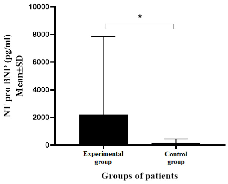
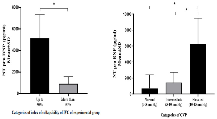
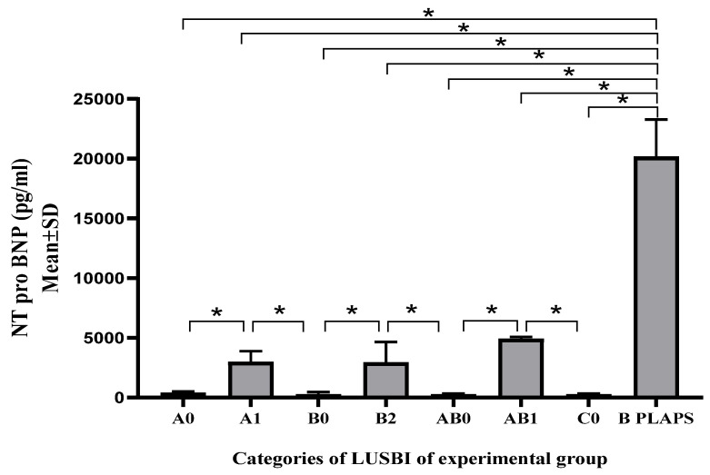
: PoCUS ultrasound applications are widely used in everyday work, especially in the field of emergency medicine. The main goal of this research was to create a diagnostic and therapeutic protocol that will integrate ultrasound examination of the lungs, ultrasound measurements of the inferior vena cava (assessment of central venous pressure) and BREST scores (risk stratification for heart failure), with the aim of establishing a more effective differential diagnostic approach for dyspneic patients. A cross-sectional study was conducted in the emergency medicine department with the educational center of the community health center of Banja Luka. Eighty patients of both sexes were included and divided into experimental and control groups based on the presence or absence of dyspnea as a dominant subjective complaint. Based on the abovementioned variables, the LUSBI protocol (lung ultrasound/BREST score/inferior vena cava) was created, including profiles to determine the nature of the origin of complaints. The biochemical marker of heart failure NT pro-BNP served as a laboratory confirmation of the cardiac origin of the complaints. : The distribution of NT pro BNP values in the experimental group showed statistically significant differences between individual profiles of the LUSBI protocol ( < 0.001). Patients assigned to group B PLAPS 2 had significantly higher average values of NT pro-BNP (20159.00 ± 3114.02 pg/mL) compared to other LUSBI profiles. Patients from the experimental group who had a high risk of heart failure according to their BREST scores also had a significantly higher average maximum expiratory diameter compared to those without heart failure ( = 0.004). A statistically significant difference ( = 0.001) in LUSBI profiles was observed between the groups of patients divided according to CVP categories. : The integration of the LUSBI protocol into the differential diagnosis of dyspnea has been shown to be very effective in confirming or excluding a cardiac cause of the disease in patients.

摘要

: 肺部超声、下腔静脉超声测量(中心静脉压评估)和 BREST 评分(心力衰竭风险分层)在日常工作中得到广泛应用,特别是在急诊医学领域。本研究的主要目的是制定一种诊断和治疗方案,将肺部超声检查、下腔静脉超声测量和 BREST 评分整合在一起,旨在为呼吸困难患者建立更有效的鉴别诊断方法。本研究为横断面研究,在巴尼亚卢卡社区卫生中心的急诊医学系和教育中心进行。共纳入 80 例男女患者,根据是否存在呼吸困难这一主要主诉,将其分为实验组和对照组。基于上述变量,制定了 LUSBI 方案(肺部超声/BREST 评分/下腔静脉),包括确定症状起源性质的特征。心力衰竭的生物标志物 NT pro-BNP 作为心脏来源症状的实验室确认。: 实验组 NT pro-BNP 值的分布在 LUSBI 方案的各个特征之间存在统计学差异( < 0.001)。B 组 PLAPS 2 患者的 NT pro-BNP 平均值显著高于其他 LUSBI 特征(20159.00 ± 3114.02 pg/mL)。根据 BREST 评分,被归类为高危心力衰竭的实验组患者的呼气末最大直径平均值也明显高于无心力衰竭患者( = 0.004)。根据 CVP 类别分组的患者中,LUSBI 特征的差异具有统计学意义( = 0.001)。: 将 LUSBI 方案纳入呼吸困难的鉴别诊断中,对于确认或排除患者疾病的心脏病因非常有效。

https://cdn.ncbi.nlm.nih.gov/pmc/blobs/d189/11433694/86ba8371566c/medicina-60-01521-g003.jpg
https://cdn.ncbi.nlm.nih.gov/pmc/blobs/d189/11433694/c3c7b4a0812e/medicina-60-01521-g001.jpg
https://cdn.ncbi.nlm.nih.gov/pmc/blobs/d189/11433694/7006bf4b162f/medicina-60-01521-g002.jpg
https://cdn.ncbi.nlm.nih.gov/pmc/blobs/d189/11433694/86ba8371566c/medicina-60-01521-g003.jpg
https://cdn.ncbi.nlm.nih.gov/pmc/blobs/d189/11433694/c3c7b4a0812e/medicina-60-01521-g001.jpg
https://cdn.ncbi.nlm.nih.gov/pmc/blobs/d189/11433694/7006bf4b162f/medicina-60-01521-g002.jpg
https://cdn.ncbi.nlm.nih.gov/pmc/blobs/d189/11433694/86ba8371566c/medicina-60-01521-g003.jpg

相似文献

1
The LUSBI Protocol (Lung Ultrasound/BREST Score/Inferior Vena Cava)-Its Role in a Differential Diagnostic Approach to Dyspnea of Cardiogenic and Non-Cardiogenic Origin.LUSBI 方案(肺部超声/ BREST 评分/下腔静脉)-其在心源性和非心源性呼吸困难鉴别诊断中的作用。
Medicina (Kaunas). 2024 Sep 18;60(9):1521. doi: 10.3390/medicina60091521.
2
Amino-terminal pro-B-type natriuretic peptide, inferior vena cava ultrasound, and biolectrical impedance analysis for the diagnosis of acute decompensated CHF.氨基末端前B型利钠肽、下腔静脉超声及生物电阻抗分析用于急性失代偿性心力衰竭的诊断。
Am J Emerg Med. 2016 Sep;34(9):1817-22. doi: 10.1016/j.ajem.2016.06.043. Epub 2016 Jun 14.
3
Rapid evaluation by lung-cardiac-inferior vena cava (LCI) integrated ultrasound for differentiating heart failure from pulmonary disease as the cause of acute dyspnea in the emergency setting.在急诊环境中,通过肺-心脏-下腔静脉(LCI)整合超声快速评估以鉴别心力衰竭与肺部疾病作为急性呼吸困难的病因。
Cardiovasc Ultrasound. 2012 Dec 4;10(1):49. doi: 10.1186/1476-7120-10-49.
4
The role of inferior vena cava diameter in the differential diagnosis of dyspneic patients; best sonographic measurement method?下腔静脉直径在呼吸困难患者鉴别诊断中的作用;最佳超声测量方法?
Am J Emerg Med. 2015 Mar;33(3):396-401. doi: 10.1016/j.ajem.2014.12.032. Epub 2014 Dec 26.
5
Which method is best for an early accurate diagnosis of acute heart failure? Comparison between lung ultrasound, chest X-ray and NT pro-BNP performance: a prospective study.哪种方法最有助于早期准确诊断急性心力衰竭?肺部超声、胸部 X 光和 NT pro-BNP 性能比较:一项前瞻性研究。
Intern Emerg Med. 2017 Sep;12(6):861-869. doi: 10.1007/s11739-016-1498-3. Epub 2016 Jul 11.
6
Rapid cardiothoracic ultrasound protocol for diagnosis of acute heart failure in the emergency department.急诊科快速心胸超声诊断急性心力衰竭方案。
Eur J Emerg Med. 2019 Apr;26(2):112-117. doi: 10.1097/MEJ.0000000000000499.
7
Correlation of natriuretic peptides and inferior vena cava size in patients with congestive heart failure.充血性心力衰竭患者利钠肽与下腔静脉大小的相关性
Vasc Health Risk Manag. 2012;8:213-8. doi: 10.2147/VHRM.S30001. Epub 2012 Apr 10.
8
Combination of lung ultrasound (a comet-tail sign) and N-terminal pro-brain natriuretic peptide in differentiating acute heart failure from chronic obstructive pulmonary disease and asthma as cause of acute dyspnea in prehospital emergency setting.在院前急救环境中,肺超声(彗尾征)与 N 末端脑利钠肽前体联合应用,有助于鉴别急性心源性呼吸困难与慢性阻塞性肺疾病和哮喘。
Crit Care. 2011;15(2):R114. doi: 10.1186/cc10140. Epub 2011 Apr 14.
9
Diagnostic accuracy of inferior vena cava evaluation in the diagnosis of acute heart failure among dyspneic patients.下腔静脉评估对呼吸困难患者急性心力衰竭诊断的准确性。
Monaldi Arch Chest Dis. 2020 Nov 9;90(4). doi: 10.4081/monaldi.2020.1375.
10
Diagnosing heart failure among acutely dyspneic patients with cardiac, inferior vena cava, and lung ultrasonography.应用心脏、下腔静脉及肺部超声诊断急性呼吸困难伴心力衰竭。
Am J Emerg Med. 2013 Aug;31(8):1208-14. doi: 10.1016/j.ajem.2013.05.007. Epub 2013 Jun 13.

本文引用的文献

1
New International Guidelines and Consensus on the Use of Lung Ultrasound.国际新指南与共识:肺超声的应用
J Ultrasound Med. 2023 Feb;42(2):309-344. doi: 10.1002/jum.16088. Epub 2022 Aug 22.
2
Ultrasound imaging and central venous pressure in spontaneously breathing patients: a comparison of ultrasound-based measures of internal jugular vein and inferior vena cava.超声成像与自主呼吸患者中心静脉压:基于超声的颈内静脉和下腔静脉测量指标的比较。
Anaesthesiol Intensive Ther. 2022;54(2):150-155. doi: 10.5114/ait.2022.114469.
3
Bedside lung ultrasonography by emergency department residents as an aid for identifying heart failure in patients with acute dyspnea after a 2-h training course.
急诊科住院医师在经过2小时培训课程后,进行床旁肺部超声检查以辅助识别急性呼吸困难患者的心力衰竭。
Ultrasound J. 2021 Feb 9;13(1):5. doi: 10.1186/s13089-021-00207-9.
4
The value of the inferior vena cava ultrasound in the decision to hospitalise in patients with acute decompensated heart failure; the best sonographic measurement method?下腔静脉超声在急性失代偿性心力衰竭患者住院决策中的价值;最佳的超声测量方法?
Acta Cardiol. 2021 May;76(3):245-257. doi: 10.1080/00015385.2020.1740422. Epub 2020 Mar 19.
5
Pulmonary Ultrasound in Patients with Heart Failure - Systematic Review.心力衰竭患者的肺部超声——系统评价
Arq Bras Cardiol. 2018 Jun;110(6):577-584. doi: 10.5935/abc.20180097.
6
Pre-hospital lung ultrasound for cardiac heart failure and COPD: is it worthwhile?院前肺部超声检查对心力衰竭和慢性阻塞性肺疾病的诊断:是否值得?
Crit Ultrasound J. 2018 Sep 10;10(1):22. doi: 10.1186/s13089-018-0104-5.
7
NT-proBNP test with improved accuracy for the diagnosis of chronic heart failure.用于慢性心力衰竭诊断的准确性有所提高的N末端B型利钠肽原检测。
Medicine (Baltimore). 2017 Dec;96(51):e9181. doi: 10.1097/MD.0000000000009181.
8
Approach to Acute Heart Failure in the Emergency Department.急诊科急性心力衰竭的处理方法
Prog Cardiovasc Dis. 2017 Sep-Oct;60(2):178-186. doi: 10.1016/j.pcad.2017.08.008. Epub 2017 Sep 1.
9
Development of a clinical prediction score for congestive heart failure diagnosis in the emergency care setting: The Brest score.用于急诊环境中充血性心力衰竭诊断的临床预测评分系统的开发:布雷斯特评分
Am J Emerg Med. 2016 Dec;34(12):2277-2283. doi: 10.1016/j.ajem.2016.08.023. Epub 2016 Aug 14.
10
The efficacy of sonographic measurement of inferior vena cava diameter as an estimate of central venous pressure.超声测量下腔静脉直径作为中心静脉压评估指标的有效性。
Cardiovasc Ultrasound. 2016 Aug 20;14(1):33. doi: 10.1186/s12947-016-0076-1.