Agapitou Chrysa, Sergentanis Theodoros N, Thymis John, Pavlidis George, Lampsas Stamatios, Korakas Emmanouil, Kountouri Aikaterini, Pliouta Loukia, Karmiris Efthymios, Lagiou Areti, Theodossiadis Panagiotis, Lambadiari Vaia, Ikonomidis Ignatios, Chatziralli Irini
2nd Department of Ophthalmology, Attikon Hospital, Medical School, National and Kapodistrian University of Athens, 124 62 Athens, Greece.
Department of Public Health Policy, School of Public Health, University of West Attica, 115 21 Athens, Greece.
J Pers Med. 2024 Sep 19;14(9):995. doi: 10.3390/jpm14090995.
To evaluate the potential association between endothelial glycocalyx damage, as well as arterial stiffness, and the retinal changes on optical coherence tomography (OCT) and OCT-angiography (OCT-A) in patients with type 2 diabetes mellitus (DM).
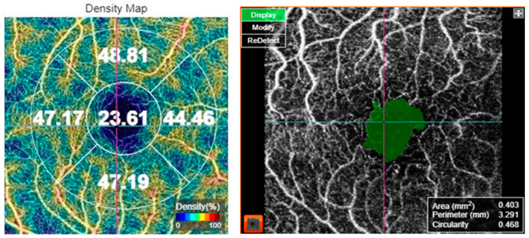
Participants in this cross-sectional study were 65 patients with DM type 2 and 42 age- and gender-matched controls without DM. The demographic and clinical characteristics of the participants were recorded. All patients underwent a thorough ophthalmological examination and multimodal imaging, including fundus photography, OCT, and OCT-A. In addition, evaluation of the endothelial glycocalyx thickness by measuring the perfused boundary region (PBR5-25) of the sublingual microvessel, as well as of the arterial stiffness, by measuring the carotid-femoral pulse wave velocity (PWV), the central aortic pressures and the augmentation index (Aix) was performed. Univariate and multivariate logistic regression analysis was performed for the examination of the potential association between the eye imaging variables and the cardiovascular-related variables. The odds ratios (OR) with the respective 95% confidence intervals (CI) were calculated. A -value < 0.05 was considered statistically significant.
Patients with DM presented significantly higher PBR5-25 compared to controls without DM ( = 0.023). At the univariate analysis, increased PBR5-25 (≥2.19 μm vs. <2.19 μm) was associated with decreased peripapillary VD at the superior quadrant (univariate OR (95% CI) = 0.34 (0.12-0.93), = 0.037). Multivariate logistic regression analysis showed that increased PWV (≥13.7 m/s vs. <13.7 m/s) was associated with an increased foveal avascular zone (FAZ) area on OCT-A ( = 0.044) and increased FAZ perimeter ( = 0.048). Moreover, increased Aix (≥14.745% vs. <14.745%) was associated with diabetic macular edema (DME) presence ( = 0.050) and increased perifoveal and parafoveal superior and temporal thickness on OCT ( < 0.05 for all associations).
Markers of endothelial damage and arterial stiffness were associated with structural and microvascular retinal alterations in patients with DM, pointing out that OCT-A could be a useful biomarker for detecting potential cardiovascular risk in such patients.
评估2型糖尿病(DM)患者内皮糖萼损伤、动脉僵硬度与光学相干断层扫描(OCT)及OCT血管造影(OCT-A)检查的视网膜变化之间的潜在关联。
本横断面研究的参与者包括65例2型糖尿病患者和42例年龄及性别匹配的非糖尿病对照者。记录参与者的人口统计学和临床特征。所有患者均接受了全面的眼科检查和多模态成像,包括眼底照相、OCT和OCT-A。此外,通过测量舌下微血管的灌注边界区域(PBR5-25)评估内皮糖萼厚度,并通过测量颈股脉搏波速度(PWV)、中心主动脉压和增强指数(Aix)评估动脉僵硬度。对眼部成像变量与心血管相关变量之间的潜在关联进行单因素和多因素逻辑回归分析。计算相应的95%置信区间(CI)的比值比(OR)。P值<0.05被认为具有统计学意义。
与非糖尿病对照者相比,糖尿病患者的PBR5-25显著更高(P = 0.023)。在单因素分析中,PBR5-25增加(≥2.19μm vs.<2.19μm)与上象限视乳头周围血管密度(VD)降低相关(单因素OR(95%CI)= 0.34(0.12 - 0.93),P = 0.037)。多因素逻辑回归分析显示,PWV增加(≥13.7 m/s vs.<13.7 m/s)与OCT-A上的黄斑无血管区(FAZ)面积增加相关(P = 0.044)以及FAZ周长增加相关(P = 0.048)。此外,Aix增加(≥14.745% vs.<14.745%)与糖尿病性黄斑水肿(DME)的存在相关(P = 0.050)以及与OCT上黄斑中心凹周围及旁中心凹上颞侧厚度增加相关(所有关联P<0.05)。
内皮损伤和动脉僵硬度标志物与糖尿病患者视网膜结构和微血管改变相关,表明OCT-A可能是检测此类患者潜在心血管风险的有用生物标志物。