Dacosta-Aguayo Rosalia, Torán-Monserrat Pere, Carmona-Cervelló Meritxell, León-Gómez Brenda Biaani, Mataró Maria, Puig Josep, Monté-Rubio Gemma, López-Lifante Victor M, Maria Manresa-Domínguez Josep, Zamora-Putin Valeria, Montero-Alia Pilar, Chacón Carla, Bielsa-Pascual Jofre, Moreno-Gabriel Eduard, García-Sierra Rosa, Rodríguez-Pérez M Carmen, Costa-Garrido Anna, Prado Julia G, Martínez-Cáceres Eva, Mateu Lourdes, Massanella Marta, Violán Concepción, Lamonja-Vicente Noemí
Unitat de Suport a la Recerca Metropolitana Nord, Institut Universitari d'Investigació en Atenció Primària Jordi Gol (IDIAP Jordi Gol), Mataró, Spain.
Germans Trias i Pujol Research Institute (IGTP), Badalona, Spain.
Front Neurol. 2024 Sep 13;15:1426881. doi: 10.3389/fneur.2024.1426881. eCollection 2024.
There is a growing interest in the effect of Long-COVID (LC) on cognition, and neuroimaging allows us to gain insight into the structural and functional changes underlying cognitive impairment in LC. We used multimodal neuroimaging data in combination with neuropsychological evaluations to study cognitive complaints in a cohort of LC patients with mild to moderate severity symptoms.
We conducted a 3T brain magnetic resonance imaging (MRI) study with diffusion tensor imaging (DTI) and functional MRI (fMRI) sequences on 53 LC patients 1.8 years after acute COVID-19 onset. We administered neuropsychological tests to evaluate cognitive domains and examined correlations with Tract-Based Spatial Statistics (TBSS) and resting state.
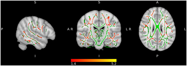
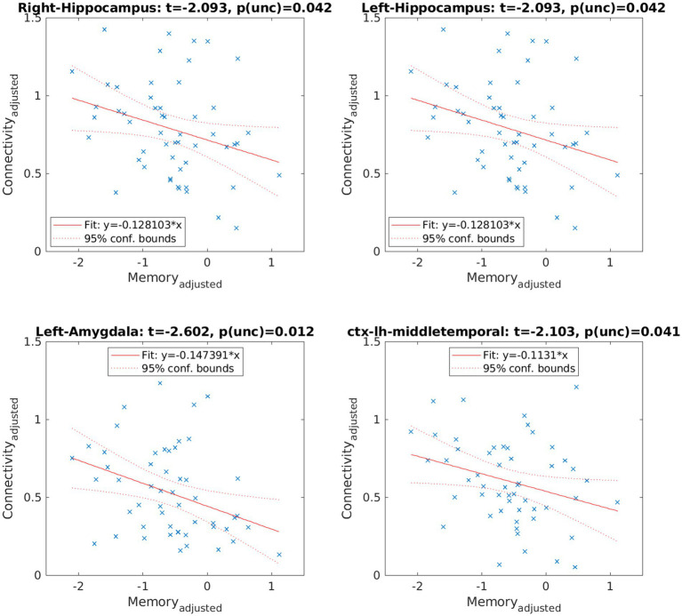
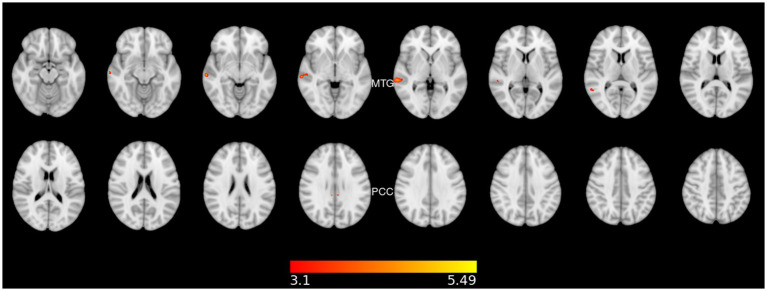
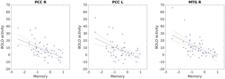
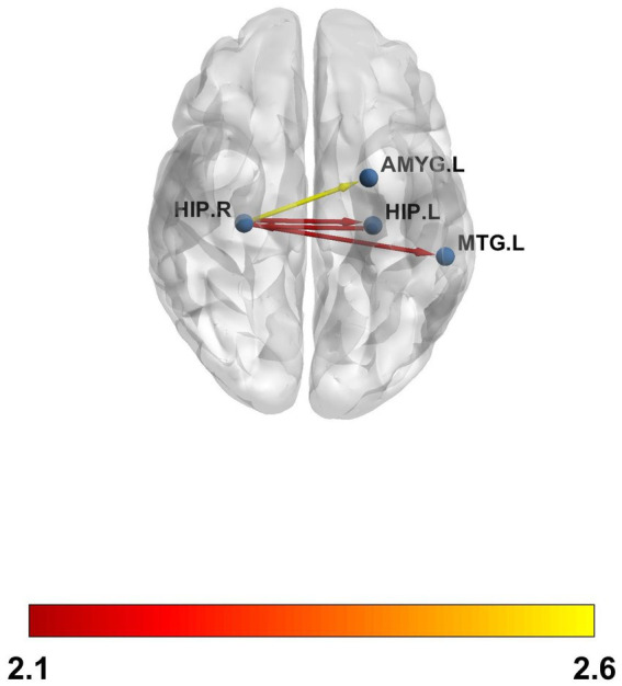
We included 53 participants with LC (mean age, 48.23 years; 88.7% females). According to the Frascati criteria, more than half of the participants had deficits in the executive (59%) and attentional (55%) domains, while 40% had impairments in the memory domain. Only one participant (1.89%) showed problems in the visuospatial and visuoconstructive domain. We observed that increased radial diffusivity in different white matter tracts was negatively correlated with the memory domain. Our results showed that higher resting state activity in the fronto-parietal network was associated with lower memory performance. Moreover, we detected increased functional connectivity among the bilateral hippocampus, the right hippocampus and the left amygdala, and the right hippocampus and the left middle temporal gyrus. These connectivity patterns were inversely related to memory and did not survive false discovery rate (FDR) correction.
People with LC exhibit cognitive impairments linked to long-lasting changes in brain structure and function, which justify the cognitive alterations detected.
人们对长新冠(LC)对认知的影响越来越感兴趣,神经影像学使我们能够深入了解LC认知障碍背后的结构和功能变化。我们使用多模态神经影像学数据结合神经心理学评估,研究了一组症状为轻至中度的LC患者的认知主诉。
我们对53名LC患者在急性新冠病毒病发作1.8年后进行了3T脑磁共振成像(MRI)研究,采用扩散张量成像(DTI)和功能MRI(fMRI)序列。我们进行了神经心理学测试以评估认知领域,并检查了与基于体素的空间统计(TBSS)和静息状态的相关性。
我们纳入了53名LC参与者(平均年龄48.23岁;88.7%为女性)。根据弗拉斯卡蒂标准,超过一半的参与者在执行功能(59%)和注意力(55%)领域存在缺陷,而40%在记忆领域存在损害。只有一名参与者(1.89%)在视觉空间和视觉构建领域存在问题。我们观察到不同白质束中径向扩散率的增加与记忆领域呈负相关。我们的结果表明,额顶叶网络中较高的静息状态活动与较低的记忆表现相关。此外,我们检测到双侧海马体、右侧海马体与左侧杏仁核以及右侧海马体与左侧颞中回之间的功能连接增加。这些连接模式与记忆呈负相关,且在错误发现率(FDR)校正后不显著。
LC患者表现出与脑结构和功能的长期变化相关的认知障碍,这证明了所检测到的认知改变是合理的。