Knoll Christoph, Doehler Juliane, Northall Alicia, Schreiber Stefanie, Rotta Johanna, Mattern Hendrik, Kuehn Esther
Institute of Cognitive Neurology and Dementia Research (IKND), Otto von Guericke University Magdeburg, Magdeburg 39120, Germany.
German Center for Neurodegenerative Diseases (DZNE) Magdeburg, Magdeburg 39120, Germany.
Brain Commun. 2024 Sep 19;6(5):fcae321. doi: 10.1093/braincomms/fcae321. eCollection 2024.
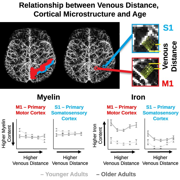
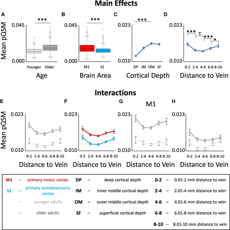
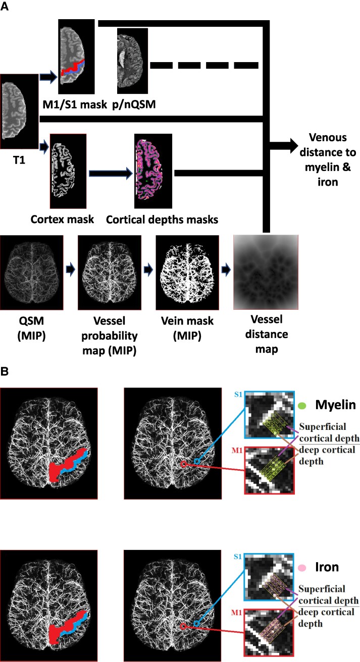
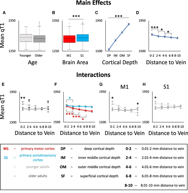
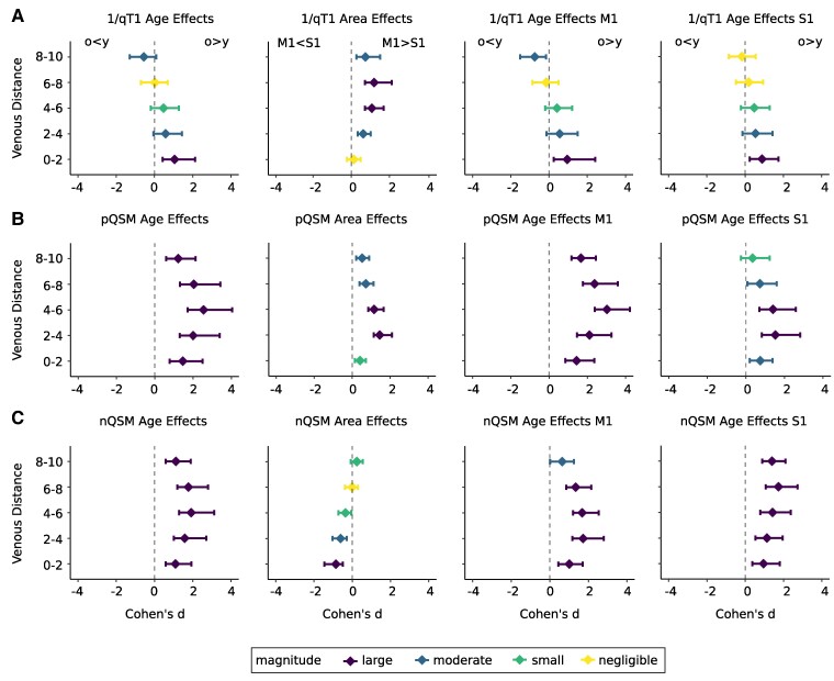
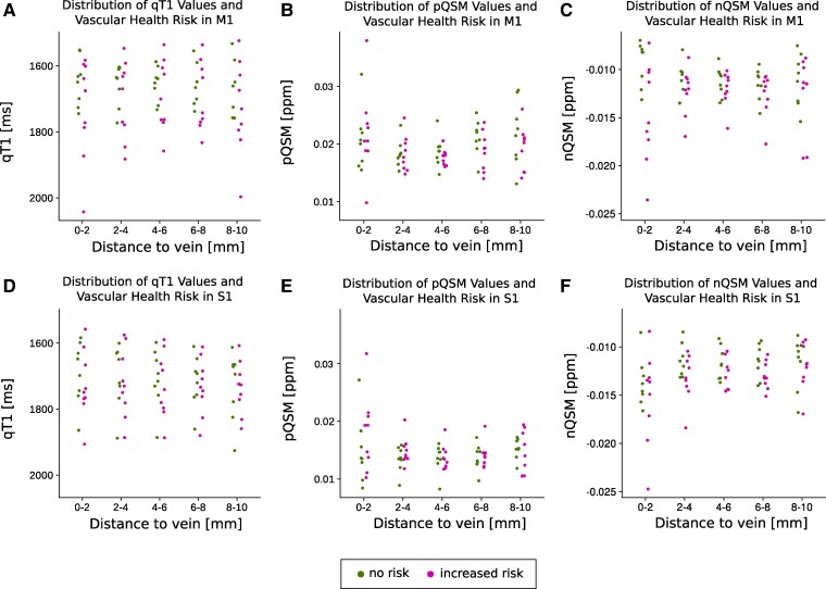
Age-related differences in cortical microstructure are used to understand the neuronal mechanisms that underlie human brain ageing. The cerebral vasculature contributes to cortical ageing, but its precise interaction with cortical microstructure is poorly understood. In a cross-sectional study, we combine venous imaging with vessel distance mapping to investigate the interaction between venous distances and age-related differences in the microstructural architecture of the primary somatosensory cortex, the primary motor cortex and additional areas in the frontal cortex as non-sensorimotor control regions. We scanned 18 younger adults and 17 older adults using 7 Tesla MRI to measure age-related changes in longitudinal relaxation time (T1) and quantitative susceptibility mapping (QSM) values at 0.5 mm isotropic resolution. We modelled different cortical depths using an equi-volume approach and assessed the distance of each voxel to its nearest vein using vessel distance mapping. Our data reveal a dependence of cortical quantitative T1 values and positive QSM values on venous distance. In addition, there is an interaction between venous distance and age on quantitative T1 values, driven by lower quantitative T1 values in older compared to younger adults in voxels that are closer to a vein. Together, our data show that the local venous architecture explains a significant amount of variance in standard measures of cortical microstructure and should be considered in neurobiological models of human brain organisation and cortical ageing.
与年龄相关的皮质微结构差异被用于理解人类大脑衰老背后的神经元机制。脑血管系统对皮质衰老有影响,但其与皮质微结构的确切相互作用却知之甚少。在一项横断面研究中,我们将静脉成像与血管距离映射相结合,以研究静脉距离与初级体感皮层、初级运动皮层以及额叶皮质其他区域(作为非感觉运动控制区域)微结构架构中与年龄相关的差异之间的相互作用。我们使用7特斯拉磁共振成像扫描了18名年轻成年人和17名年长成年人,以测量在0.5毫米各向同性分辨率下纵向弛豫时间(T1)和定量磁化率映射(QSM)值的年龄相关变化。我们使用等体积方法对不同的皮质深度进行建模,并使用血管距离映射评估每个体素到其最近静脉的距离。我们的数据揭示了皮质定量T1值和正QSM值对静脉距离的依赖性。此外,在靠近静脉的体素中,年长成年人的定量T1值低于年轻成年人,这导致静脉距离和年龄在定量T1值上存在相互作用。总之,我们的数据表明,局部静脉结构解释了皮质微结构标准测量中相当大的方差,在人类脑组织和皮质衰老的神经生物学模型中应予以考虑。