• 文献检索
  • 文档翻译
  • 深度研究
  • 学术资讯
  • Suppr Zotero 插件Zotero 插件
  • 邀请有礼
  • 套餐&价格
  • 历史记录
应用&插件
Suppr Zotero 插件Zotero 插件浏览器插件Mac 客户端Windows 客户端微信小程序
定价
高级版会员购买积分包购买API积分包
服务
文献检索文档翻译深度研究API 文档MCP 服务
关于我们
关于 Suppr公司介绍联系我们用户协议隐私条款
关注我们

Suppr 超能文献

核心技术专利:CN118964589B侵权必究
粤ICP备2023148730 号-1Suppr @ 2026

文献检索

告别复杂PubMed语法,用中文像聊天一样搜索,搜遍4000万医学文献。AI智能推荐,让科研检索更轻松。

立即免费搜索

文件翻译

保留排版,准确专业,支持PDF/Word/PPT等文件格式,支持 12+语言互译。

免费翻译文档

深度研究

AI帮你快速写综述,25分钟生成高质量综述,智能提取关键信息,辅助科研写作。

立即免费体验

光学相干断层扫描血管造影在青光眼中的应用:现状与未来方向

Applications of optical coherence tomography angiography in glaucoma: current status and future directions.

作者信息

Shen Ruyue, Chan Leo Ka Yu, Yip Amber Cheuk Wing, Chan Poemen P

机构信息

Department of Ophthalmology and Visual Sciences, The Chinese University of Hong Kong, Hong Kong, China.

Jet King-Shing Ho Glaucoma Treatment and Research Centre, Department of Ophthalmology and Visual Sciences, The Chinese University of Hong Kong, Hong Kong, China.

出版信息

Front Med (Lausanne). 2024 Sep 19;11:1428850. doi: 10.3389/fmed.2024.1428850. eCollection 2024.

DOI:10.3389/fmed.2024.1428850
PMID:39364027
原文链接:https://pmc.ncbi.nlm.nih.gov/articles/PMC11446750/
Abstract

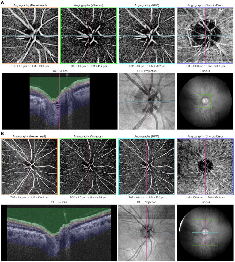
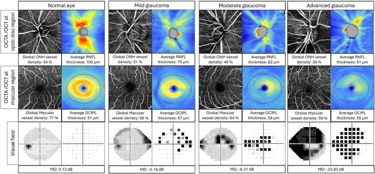
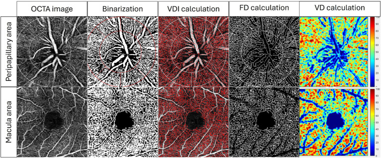
Glaucoma is a leading cause of irreversible blindness worldwide, with its pathophysiology remaining inadequately understood. Among the various proposed theories, the vascular theory, suggesting a crucial role of retinal vasculature deterioration in glaucoma onset and progression, has gained significant attention. Traditional imaging techniques, such as fundus fluorescein angiography, are limited by their invasive nature, time consumption, and qualitative output, which restrict their efficacy in detailed retinal vessel examination. Optical coherence tomography angiography (OCTA) emerges as a revolutionary imaging modality, offering non-invasive, detailed visualization of the retinal and optic nerve head microvasculature, thereby marking a significant advancement in glaucoma diagnostics and management. Since its introduction, OCTA has been extensively utilized for retinal vasculature imaging, underscoring its potential to enhance our understanding of glaucoma's pathophysiology, improving diagnosis, and monitoring disease progression. This review aims to summarize the current knowledge regarding the role of OCTA in glaucoma, particularly its potential applications in diagnosing, monitoring, and understanding the pathophysiology of the disease. Parameters pertinent to glaucoma will be elucidated to illustrate the utility of OCTA as a tool to guide glaucoma management.

摘要

青光眼是全球不可逆性失明的主要原因之一,其病理生理学仍未得到充分理解。在各种提出的理论中,血管学说认为视网膜血管病变在青光眼的发生和发展中起关键作用,该学说已引起了广泛关注。传统的成像技术,如眼底荧光血管造影,因其具有侵入性、耗时且输出结果定性等局限性,限制了它们在详细检查视网膜血管方面的功效。光学相干断层扫描血管造影(OCTA)作为一种革命性的成像方式应运而生,它能够对视网膜和视神经乳头微血管进行非侵入性的详细可视化,从而在青光眼的诊断和管理方面取得了重大进展。自问世以来,OCTA已被广泛用于视网膜血管成像,凸显了其在增进我们对青光眼病理生理学的理解、改善诊断以及监测疾病进展方面的潜力。本综述旨在总结当前关于OCTA在青光眼中作用的知识,特别是其在诊断、监测和理解该疾病病理生理学方面的潜在应用。将阐明与青光眼相关的参数,以说明OCTA作为指导青光眼管理工具的实用性。

https://cdn.ncbi.nlm.nih.gov/pmc/blobs/4ed9/11446750/e3bbc67913da/fmed-11-1428850-g004.jpg
https://cdn.ncbi.nlm.nih.gov/pmc/blobs/4ed9/11446750/948e751af550/fmed-11-1428850-g001.jpg
https://cdn.ncbi.nlm.nih.gov/pmc/blobs/4ed9/11446750/7edc29b50838/fmed-11-1428850-g002.jpg
https://cdn.ncbi.nlm.nih.gov/pmc/blobs/4ed9/11446750/41373e24ef90/fmed-11-1428850-g003.jpg
https://cdn.ncbi.nlm.nih.gov/pmc/blobs/4ed9/11446750/e3bbc67913da/fmed-11-1428850-g004.jpg

相似文献

1
Applications of optical coherence tomography angiography in glaucoma: current status and future directions.光学相干断层扫描血管造影在青光眼中的应用:现状与未来方向
Front Med (Lausanne). 2024 Sep 19;11:1428850. doi: 10.3389/fmed.2024.1428850. eCollection 2024.
2
Optical Coherence Tomography Angiography in Glaucoma Care.光学相干断层扫描血管造影在青光眼治疗中的应用。
Curr Eye Res. 2018 Sep;43(9):1067-1082. doi: 10.1080/02713683.2018.1475013. Epub 2018 May 23.
3
Optical Coherence Tomography and Optical Coherence Tomography Angiography: Essential Tools for Detecting Glaucoma and Disease Progression.光学相干断层扫描和光学相干断层扫描血管造影:检测青光眼和疾病进展的重要工具。
Front Ophthalmol (Lausanne). 2023;3. doi: 10.3389/fopht.2023.1217125. Epub 2023 Jul 28.
4
The Role of Optical Coherence Tomography Angiography in Glaucoma.光学相干断层扫描血管造影在青光眼中的作用。
Semin Ophthalmol. 2024 Aug;39(6):412-423. doi: 10.1080/08820538.2024.2343049. Epub 2024 Apr 20.
5
Optical coherence tomography angiography: an overview of the technology and an assessment of applications for clinical research.光学相干断层扫描血管造影术:技术概述及临床研究应用评估
Br J Ophthalmol. 2017 Jan;101(1):16-20. doi: 10.1136/bjophthalmol-2016-309389. Epub 2016 Oct 4.
6
Potential applications of optical coherence tomography angiography in glaucoma.光学相干断层扫描血管造影术在青光眼方面的潜在应用
Curr Opin Ophthalmol. 2018 May;29(3):226-233. doi: 10.1097/ICU.0000000000000475.
7
A comprehensive update on the use of optical coherence tomography angiography in glaucoma.光学相干断层扫描血管造影在青光眼应用的综合更新。
Int Ophthalmol. 2023 May;43(5):1785-1802. doi: 10.1007/s10792-022-02574-1. Epub 2022 Dec 6.
8
Optical Coherence Tomography Angiography in Glaucoma.光学相干断层扫描血管造影术在青光眼中的应用。
J Glaucoma. 2020 Apr;29(4):312-321. doi: 10.1097/IJG.0000000000001463.
9
[Optical coherence tomography angiography: Value for glaucoma diagnostics].[光学相干断层扫描血管造影术:在青光眼诊断中的价值]
Ophthalmologe. 2019 Jul;116(7):602-609. doi: 10.1007/s00347-018-0815-9.
10
Optical Coherence Tomography Angiography in Retinal Vascular Disorders.视网膜血管疾病中的光学相干断层扫描血管造影术
Diagnostics (Basel). 2023 May 3;13(9):1620. doi: 10.3390/diagnostics13091620.

本文引用的文献

1
Diagnostic performance of wide-field optical coherence tomography angiography for high myopic glaucoma.广角光学相干断层扫描血管造影术在高度近视性青光眼诊断中的性能。
Sci Rep. 2024 Jan 3;14(1):367. doi: 10.1038/s41598-023-49542-y.
2
Peripapillary Perfusion Analysis Using Optical Coherence Tomography Angiography in Patients with Normal Tension Glaucoma.用光学相干断层扫描血管造影术分析正常眼压青光眼患者的视盘周围灌注。
Korean J Ophthalmol. 2024 Feb;38(1):51-63. doi: 10.3341/kjo.2023.0112. Epub 2023 Dec 19.
3
Advances in swept-source optical coherence tomography and optical coherence tomography angiography.
扫频源光学相干断层扫描及光学相干断层扫描血管造影术的进展。
Adv Ophthalmol Pract Res. 2022 Nov 25;3(2):67-79. doi: 10.1016/j.aopr.2022.10.005. eCollection 2023 May-Jun.
4
Temporal Optic Disc Microvasculature Dropout in Glaucoma.青光眼的视盘微血管时间性缺失。
Invest Ophthalmol Vis Sci. 2023 Aug 1;64(11):6. doi: 10.1167/iovs.64.11.6.
5
Optical coherence tomography angiography: a review of the current literature.光学相干断层扫描血管造影:当前文献综述。
J Int Med Res. 2023 Jul;51(7):3000605231187933. doi: 10.1177/03000605231187933.
6
Association of superficial macular vessel density with visual field progression in open-angle glaucoma with central visual field damage.在伴有中央视野损害的开角型青光眼患者中,浅层视网膜血管密度与视野进展的相关性。
Sci Rep. 2023 May 3;13(1):7190. doi: 10.1038/s41598-023-34000-6.
7
Normal tension glaucoma: A dynamic optical coherence tomography angiography study.正常眼压性青光眼:一项动态光学相干断层扫描血管造影研究。
Front Med (Lausanne). 2023 Jan 6;9:1037471. doi: 10.3389/fmed.2022.1037471. eCollection 2022.
8
Ultra-Widefield OCT Angiography.超广角 OCT 血管造影。
IEEE Trans Med Imaging. 2023 Apr;42(4):1009-1020. doi: 10.1109/TMI.2022.3222638. Epub 2023 Apr 3.
9
A standardized method to quantitatively analyze optical coherence tomography angiography images of the macular and peripapillary vessels.一种定量分析黄斑和视乳头周围血管光学相干断层扫描血管造影图像的标准化方法。
Int J Retina Vitreous. 2022 Oct 15;8(1):75. doi: 10.1186/s40942-022-00426-9.
10
Fractal Dimension of Peripapillary Vasculature in Primary Open-Angle Glaucoma.原发性开角型青光眼的视盘周围血管分形维数。
Korean J Ophthalmol. 2022 Dec;36(6):518-526. doi: 10.3341/kjo.2022.0089. Epub 2022 Oct 11.