• 文献检索
  • 文档翻译
  • 深度研究
  • 学术资讯
  • Suppr Zotero 插件Zotero 插件
  • 邀请有礼
  • 套餐&价格
  • 历史记录
应用&插件
Suppr Zotero 插件Zotero 插件浏览器插件Mac 客户端Windows 客户端微信小程序
定价
高级版会员购买积分包购买API积分包
服务
文献检索文档翻译深度研究API 文档MCP 服务
关于我们
关于 Suppr公司介绍联系我们用户协议隐私条款
关注我们

Suppr 超能文献

核心技术专利:CN118964589B侵权必究
粤ICP备2023148730 号-1Suppr @ 2026

文献检索

告别复杂PubMed语法,用中文像聊天一样搜索,搜遍4000万医学文献。AI智能推荐,让科研检索更轻松。

立即免费搜索

文件翻译

保留排版,准确专业,支持PDF/Word/PPT等文件格式,支持 12+语言互译。

免费翻译文档

深度研究

AI帮你快速写综述,25分钟生成高质量综述,智能提取关键信息,辅助科研写作。

立即免费体验

MRI 影像学生物标志物在颅内压测定中的应用。

The utility of MRI radiological biomarkers in determining intracranial pressure.

机构信息

High-Dimensional Neurology Group, UCL Queen Square Institute of Neurology, University College London, London, UK.

Victor Horsley Department of Neurosurgery, National Hospital for Neurology and Neurosurgery, London, UK.

出版信息

Sci Rep. 2024 Oct 5;14(1):23238. doi: 10.1038/s41598-024-73750-9.

DOI:10.1038/s41598-024-73750-9
PMID:39369053
原文链接:https://pmc.ncbi.nlm.nih.gov/articles/PMC11455934/
Abstract

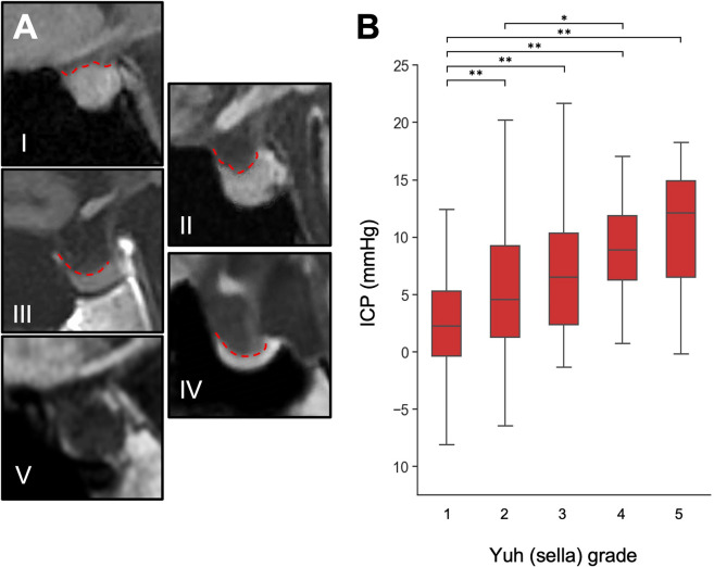
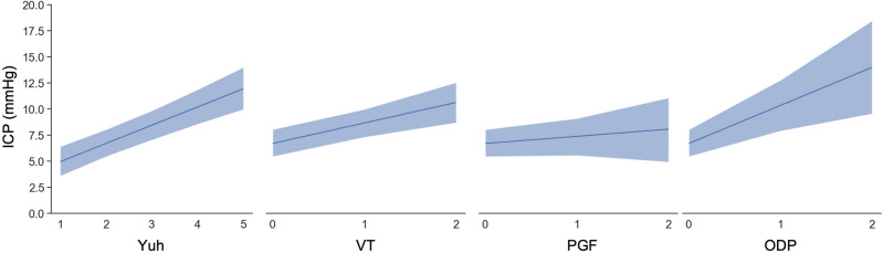
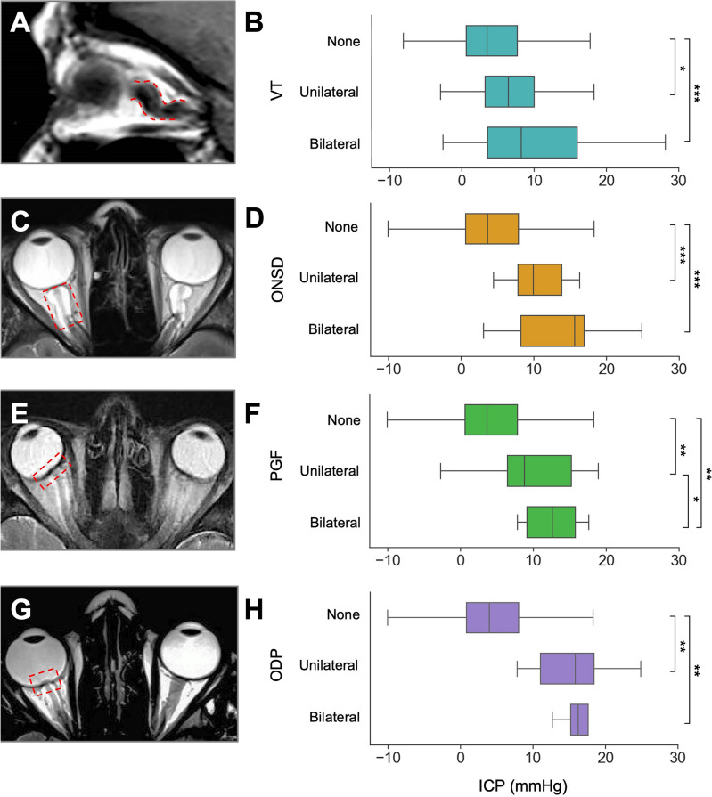
Intracranial pressure (ICP) is a physiological parameter that conventionally requires invasive monitoring for accurate measurement. Utilising multivariate predictive models, we sought to evaluate the utility of non-invasive, widely accessible MRI biomarkers in predicting ICP and their reversibility following cerebrospinal fluid (CSF) diversion. The retrospective study included 325 adult patients with suspected CSF dynamic disorders who underwent brain MRI scans within three months of elective 24-h ICP monitoring. Five MRI biomarkers were assessed: Yuh sella grade, optic nerve vertical tortuosity (VT), optic nerve sheath distension, posterior globe flattening and optic disc protrusion (ODP). The association between individual biomarkers and 24-h ICP was examined and reversibility of each following CSF diversion was assessed. Multivariate models incorporating these radiological biomarkers were utilised to predict 24-h median intracranial pressure. All five biomarkers were significantly associated with median 24-h ICP (p < 0.0001). Using a pair-wise approach, the presence of each abnormal biomarker was significantly associated with higher median 24-h ICP (p < 0.0001). On multivariate analysis, ICP was significantly and positively associated with Yuh sella grade (p < 0.0001), VT (p < 0.0001) and ODP (p = 0.003), after accounting for age and suspected diagnosis. The Bayesian multiple linear regression model predicted 24-h median ICP with a mean absolute error of 2.71 mmHg. Following CSF diversion, we found pituitary sella grade to show significant pairwise reversibility (p < 0.001). ICP was predicted with clinically useful precision utilising a compact Bayesian model, offering an easily interpretable tool using non-invasive MRI data. Brain MRI biomarkers are anticipated to play a more significant role in the screening, triaging, and referral of patients with suspected CSF dynamic disorders.

摘要

颅内压(ICP)是一种生理参数,传统上需要进行有创监测才能进行准确测量。我们利用多变量预测模型,旨在评估非侵入性、广泛可获得的 MRI 生物标志物在预测 ICP 及其在脑脊液(CSF)分流后的可逆转性方面的效用。这项回顾性研究纳入了 325 名疑似 CSF 动力学障碍的成年患者,这些患者在 24 小时 ICP 监测后的三个月内接受了脑部 MRI 扫描。评估了 5 种 MRI 生物标志物:蝶鞍 Yuh 分级、视神经垂直迂曲(VT)、视神经鞘扩张、后眼球变平及视盘突出(ODP)。检查了各个生物标志物与 24 小时 ICP 的相关性,并评估了每种生物标志物在 CSF 分流后的可逆转性。利用纳入这些影像学生物标志物的多变量模型来预测 24 小时的颅内压中位数。所有 5 种生物标志物与 24 小时 ICP 中位数均显著相关(p<0.0001)。采用两两比较的方法,每个异常生物标志物的存在均与较高的 24 小时 ICP 中位数显著相关(p<0.0001)。在多变量分析中,在考虑年龄和疑似诊断后,ICP 与蝶鞍 Yuh 分级(p<0.0001)、VT(p<0.0001)和 ODP(p=0.003)呈显著正相关。贝叶斯多元线性回归模型对 24 小时 ICP 中位数进行预测,平均绝对误差为 2.71mmHg。CSF 分流后,我们发现垂体蝶鞍分级具有显著的两两可逆转性(p<0.001)。使用紧凑的贝叶斯模型,ICP 可利用具有临床实用精度的预测,提供了一种使用非侵入性 MRI 数据的易于解释的工具。预计 MRI 生物标志物将在疑似 CSF 动力学障碍患者的筛查、分类和转诊中发挥更重要的作用。

https://cdn.ncbi.nlm.nih.gov/pmc/blobs/91af/11455934/15a7c50c388f/41598_2024_73750_Fig3_HTML.jpg
https://cdn.ncbi.nlm.nih.gov/pmc/blobs/91af/11455934/3202ac61913b/41598_2024_73750_Fig1_HTML.jpg
https://cdn.ncbi.nlm.nih.gov/pmc/blobs/91af/11455934/a8e82b2f7576/41598_2024_73750_Fig2_HTML.jpg
https://cdn.ncbi.nlm.nih.gov/pmc/blobs/91af/11455934/15a7c50c388f/41598_2024_73750_Fig3_HTML.jpg
https://cdn.ncbi.nlm.nih.gov/pmc/blobs/91af/11455934/3202ac61913b/41598_2024_73750_Fig1_HTML.jpg
https://cdn.ncbi.nlm.nih.gov/pmc/blobs/91af/11455934/a8e82b2f7576/41598_2024_73750_Fig2_HTML.jpg
https://cdn.ncbi.nlm.nih.gov/pmc/blobs/91af/11455934/15a7c50c388f/41598_2024_73750_Fig3_HTML.jpg

相似文献

1
The utility of MRI radiological biomarkers in determining intracranial pressure.MRI 影像学生物标志物在颅内压测定中的应用。
Sci Rep. 2024 Oct 5;14(1):23238. doi: 10.1038/s41598-024-73750-9.
2
Brain MRI and Ophthalmic Biomarkers of Intracranial Pressure.颅内压的脑部磁共振成像和眼科生物标志物
Neurology. 2021 May 31;96(22):e2714-e2723. doi: 10.1212/WNL.0000000000012023.
3
MRI findings as markers of idiopathic intracranial hypertension.MRI 表现作为特发性颅内高压的标志物。
Curr Opin Neurol. 2021 Feb 1;34(1):75-83. doi: 10.1097/WCO.0000000000000885.
4
Does bilateral transverse cerebral venous sinus stenosis exist in patients without increased intracranial pressure?颅内压未升高的患者是否存在双侧横窦狭窄?
Clin Neurol Neurosurg. 2013 Aug;115(8):1215-9. doi: 10.1016/j.clineuro.2012.11.004. Epub 2012 Dec 5.
5
The diagnostic value of cranial MRI findings in idiopathic intracranial hypertension: evaluating radiological parameters associated with intracranial pressure.特发性颅内高压症头颅 MRI 表现的诊断价值:评估与颅内压相关的影像学参数。
Acta Radiol. 2022 Oct;63(10):1390-1397. doi: 10.1177/02841851211038803. Epub 2021 Aug 23.
6
Use of T2-weighted magnetic resonance imaging of the optic nerve sheath to detect raised intracranial pressure.使用视神经鞘的T2加权磁共振成像检测颅内压升高。
Crit Care. 2008;12(5):R114. doi: 10.1186/cc7006. Epub 2008 Sep 11.
7
Spaceflight-Induced Visual Impairment and Globe Deformations in Astronauts Are Linked to Orbital Cerebrospinal Fluid Volume Increase.宇航员的太空飞行诱发视力损害和眼球变形与轨道脑脊液体积增加有关。
Acta Neurochir Suppl. 2018;126:215-219. doi: 10.1007/978-3-319-65798-1_44.
8
Use of neuroimaging measurements of optic nerve sheath diameter to assess intracranial pressure in craniosynostosis.利用视神经鞘直径的神经影像学测量评估颅缝早闭症中的颅内压。
Childs Nerv Syst. 2018 May;34(5):939-946. doi: 10.1007/s00381-018-3728-7. Epub 2018 Jan 29.
9
Comparison of accuracy of optic nerve ultrasound for the detection of intracranial hypertension in the setting of acutely fluctuating vs stable intracranial pressure: post-hoc analysis of data from a prospective, blinded single center study.急性波动与稳定颅内压情况下视神经超声检测颅内高压准确性的比较:一项前瞻性、单中心盲法研究数据的事后分析
Crit Care. 2012 May 11;16(3):R79. doi: 10.1186/cc11336.
10
Clinical Relevance of Transorbital Ultrasonographic Measurement of Optic Nerve Sheath Diameter (ONSD) for Estimation of Intracranial Pressure Following Cerebrospinal Fluid Diversion Surgery.经眶超声测量视神经鞘直径(ONSD)在脑脊液分流术后颅内压评估中的临床意义
Cureus. 2022 May 22;14(5):e25200. doi: 10.7759/cureus.25200. eCollection 2022 May.

本文引用的文献

1
Can clinicians correctly predict intracranial pressure state based on clinical symptoms alone in shunted patients?临床医生能否仅根据临床症状正确预测分流患者的颅内压状态?
J Neurosurg Sci. 2025 Feb;69(1):131-135. doi: 10.23736/S0390-5616.23.06065-4. Epub 2023 Nov 23.
2
Brain MRI and Ophthalmic Biomarkers of Intracranial Pressure.颅内压的脑部磁共振成像和眼科生物标志物
Neurology. 2021 May 31;96(22):e2714-e2723. doi: 10.1212/WNL.0000000000012023.
3
An Update on Imaging in Idiopathic Intracranial Hypertension.特发性颅内高压的影像学最新进展
Front Neurol. 2020 Jun 10;11:453. doi: 10.3389/fneur.2020.00453. eCollection 2020.
4
Review: pathophysiology of intracranial hypertension and noninvasive intracranial pressure monitoring.综述:颅内高压的病理生理学和无创颅内压监测。
Fluids Barriers CNS. 2020 Jun 23;17(1):40. doi: 10.1186/s12987-020-00201-8.
5
Association of Intracranial Pressure and Spontaneous Retinal Venous Pulsation.颅内压与自发性视网膜静脉搏动的关系。
JAMA Neurol. 2019 Dec 1;76(12):1502-1505. doi: 10.1001/jamaneurol.2019.2935.
6
Intraparenchymal intracranial pressure monitoring for hydrocephalus and cerebrospinal fluid disorders.脑实质内颅内压监测在脑积水和脑脊液疾病中的应用
Acta Neurochir (Wien). 2017 Oct;159(10):1967-1978. doi: 10.1007/s00701-017-3281-2. Epub 2017 Aug 10.
7
Imaging of Intracranial Pressure Disorders.颅内压紊乱的影像学检查
Neurosurgery. 2017 Mar 1;80(3):341-354. doi: 10.1227/NEU.0000000000001362.
8
Sonography of the Optic Nerve Sheath Diameter for Detection of Raised Intracranial Pressure Compared to Computed Tomography: A Systematic Review and Meta-analysis.与计算机断层扫描相比,超声测量视神经鞘直径用于检测颅内压升高的系统评价和荟萃分析
J Ultrasound Med. 2015 Jul;34(7):1285-94. doi: 10.7863/ultra.34.7.1285.
9
Intracranial Pressure Monitoring: Invasive versus Non-Invasive Methods-A Review.颅内压监测:有创与无创方法综述
Crit Care Res Pract. 2012;2012:950393. doi: 10.1155/2012/950393. Epub 2012 Jun 8.
10
MR imaging of papilledema and visual pathways: effects of increased intracranial pressure and pathophysiologic mechanisms.视乳头水肿和视觉通路的磁共振成像:颅内压增高的影响和病理生理机制。
AJNR Am J Neuroradiol. 2013 May;34(5):919-24. doi: 10.3174/ajnr.A3022. Epub 2012 Mar 15.