Pârvǎnescu Cristina Dorina, Bǎrbulescu Andreea Lili, Dinescu Ştefan Cristian, Bițǎ Cristina Elena, Firulescu Sineta Cristina, Traşcǎ Beatrice Andreea, Dascǎlu Rucsandra Cristina, Sandu Raluca Elena, Vreju Florentin Ananu
PhD Student, University of Medicine and Pharmacy of Craiova, Romania.
Department of Pharmacology, University of Medicine and Pharmacy of Craiova, Romania.
Curr Health Sci J. 2024 Apr-Jun;50(2):274-282. doi: 10.12865/CHSJ.50.02.13. Epub 2024 Jun 30.
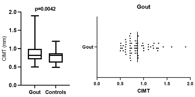
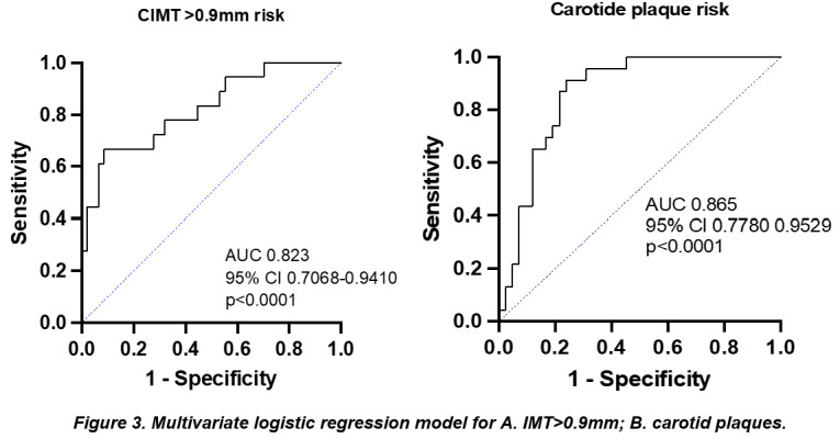
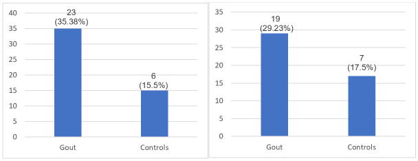
The current observational, prospective study enrolled 65 patients with gout, diagnosed according to 2015 ACR/EULAR criteria [17], evaluated in Rheumatology Clinic, Emergency County Hospital Craiova, and 40 healthy subjects. This research aimed to determine the presence of subclinical carotid atherosclerosis, revealed by an increased intima media thickness and carotid plaques in gout patients, by US examination. Secondary, we aimed to search for the possible correlations displayed between the presence of subclinical carotid atherosclerosis and several disease variables. CCAIMT over 0.9mm was identified for 19 patients (29.23%), percentage statistically significant different compared to controls (7; 17.5%), p=0,0428. For 23 patients (35.38%) carotid plaques were present at US examination, more prevalent compared to controls (19; 29.23%), p=0.002. Using multivariate logistic regression, we pointed out that SUA (OR 2,103; p=0.0002), age (OR=1,051; p<0.001), disease duration (OR=1.740; p=0.0039) and LDLc (OR=1,003; p=0.0029) were independently associated to an increased IMT in patients with gout, similar results being obtained for carotid plaques. MSKUS was performed for all patients, with important results. The presence of deposits associated with an increased risk of a thick IMT; similar results were obtained for double contour sign, aggregates and tophi. A statistically significant risk was noticed for the presence of deposits (p=0.002). Regarding the presence of carotid atheroma plaques, a higher risk was associated to deposits identification, double contour sign, aggregates, tophi and PD signal. Our results sustain that carotid ultrasound is an easily accessible imagistic method that offers important predictors of atherosclerotic status.
本项前瞻性观察性研究纳入了65例痛风患者,这些患者均根据2015年美国风湿病学会/欧洲抗风湿病联盟(ACR/EULAR)标准[17]进行诊断,在克拉约瓦县急诊医院风湿病诊所接受评估,同时纳入了40名健康受试者。本研究旨在通过超声检查确定痛风患者内膜中层厚度增加和颈动脉斑块所揭示的亚临床颈动脉粥样硬化的存在情况。其次,我们旨在探寻亚临床颈动脉粥样硬化的存在与若干疾病变量之间可能存在的相关性。19例患者(29.23%)的颈总动脉内膜中层厚度(CCAIMT)超过0.9mm,与对照组(7例;17.5%)相比,该百分比具有统计学显著差异,p = 0.0428。23例患者(35.38%)在超声检查时发现有颈动脉斑块,比对照组(19例;29.23%)更为普遍,p = 0.002。通过多因素逻辑回归分析,我们指出血尿酸(SUA)(比值比[OR]为2.103;p = 0.0002)、年龄(OR = 1.051;p < 0.001)、病程(OR = 1.740;p = 0.0039)和低密度脂蛋白胆固醇(LDLc)(OR = 1.003;p = 0.0029)与痛风患者内膜中层厚度增加独立相关,颈动脉斑块情况也得到了类似结果。对所有患者均进行了肌肉骨骼超声(MSKUS)检查,结果很重要。沉积物的存在与内膜中层厚度增厚风险增加相关;双轮廓征、聚集物和痛风石也得到了类似结果。沉积物的存在具有统计学显著风险(p = 0.002)。关于颈动脉粥样斑块的存在,沉积物识别、双轮廓征、聚集物、痛风石和功率多普勒(PD)信号与之相关的风险更高。我们的结果支持颈动脉超声是一种易于获得的成像方法,可提供动脉粥样硬化状态的重要预测指标。