Das Liza, Khadwal Alka, Malhotra Pankaj, Ghosh Jayaditya, Dhiman Vandana, Sharma Vivek, Singhmar Shallu, Ahuja Chirag Kamal, Saikia Uma Nahar, Bhadada Sanjay Kumar, Dutta Pinaki
Department of Endocrinology, Post Graduate Institute of Medical Education and Research, Chandigarh 160012, India.
Department of Telemedicine, Post Graduate Institute of Medical Education and Research, Chandigarh 160012, India.
JBMR Plus. 2024 Aug 30;8(11):ziae117. doi: 10.1093/jbmrpl/ziae117. eCollection 2024 Nov.
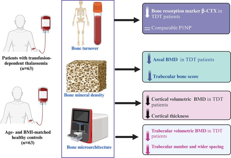
Thalassemic osteopathy includes low bone mass and impaired bone microarchitecture. We aimed to evaluate the prevalence and determinants of bone quantity (osteoporosis) and quality (microarchitecture) in a cohort of adult patients with transfusion-dependent thalassemia (TDT). Patients with TDT ( = 63) and age- and BMI-matched controls ( = 63) were recruited in the study. Areal bone mineral density (BMD) was measured using DXA Hologic scanner. P1NP and β-CTX were estimated by electrochemiluminescence assay. Bone geometry and volumetric BMD (vBMD) were estimated by second-generation high-resolution peripheral quantitative computed tomography. Bone turnover marker β-CTX was significantly lower in the TDT group, but there was no difference in P1NP levels. Low bone mass (Z ≤ -2) was present in greater proportion of patients both at lumbar spine (LS) (54 vs 0%; = .001) and femoral neck (FN) (33 vs 8%; = .001). Hypogonadism was associated with low BMD at FN (OR 10.0; 95% CI, 1.2-86; = .01) and low hemoglobin with low BMD at LS (OR 1.58; 95% CI, 0.96-2.60; = .07). The mean trabecular bone score was also significantly lower in patients compared with controls (1.261 ± 0.072 vs 1.389 ± 0.058). Total, cortical and trabecular vBMD were significantly lower in cases than controls. The trabecular number and cortical thickness were significantly lower and trabecular separation higher in cases than controls. Adults with TDT have significantly lower areal, cortical and trabecular vBMD. The bone microarchitecture is also significantly impaired in terms of lower number and wider spacing of trabeculae as well as lower cortical thickness and area at both radius and tibia.
地中海贫血性骨病包括低骨量和骨微结构受损。我们旨在评估一组依赖输血的成年地中海贫血(TDT)患者的骨量(骨质疏松症)和质量(微结构)的患病率及决定因素。本研究招募了TDT患者(n = 63)以及年龄和体重指数匹配的对照组(n = 63)。使用双能X线吸收仪(DXA)Hologic扫描仪测量面积骨密度(BMD)。通过电化学发光法测定I型前胶原氨基端前肽(P1NP)和I型胶原交联C端肽(β-CTX)。通过第二代高分辨率外周定量计算机断层扫描评估骨几何结构和体积骨密度(vBMD)。TDT组的骨转换标志物β-CTX显著降低,但P1NP水平无差异。腰椎(LS)(54% 对0%;P = .001)和股骨颈(FN)(33% 对8%;P = .001)处,骨量低(Z≤ -2)的患者比例更高。性腺功能减退与FN处低骨密度相关(比值比[OR] 10.0;95%置信区间[CI],1.2 - 86;P = .01),而低血红蛋白与LS处低骨密度相关(OR 1.58;95% CI,0.96 - 2.60;P = .07)。与对照组相比,患者的平均小梁骨评分也显著更低(1.261 ± 0.072对1.389 ± 0.058)。病例组的总体、皮质和小梁vBMD显著低于对照组。病例组的小梁数量和皮质厚度显著更低,小梁间距更高。成年TDT患者的面积、皮质和小梁vBMD显著更低。在小梁数量减少、间距增宽以及桡骨和胫骨的皮质厚度和面积降低方面,骨微结构也显著受损。