• 文献检索
  • 文档翻译
  • 深度研究
  • 学术资讯
  • Suppr Zotero 插件Zotero 插件
  • 邀请有礼
  • 套餐&价格
  • 历史记录
应用&插件
Suppr Zotero 插件Zotero 插件浏览器插件Mac 客户端Windows 客户端微信小程序
定价
高级版会员购买积分包购买API积分包
服务
文献检索文档翻译深度研究API 文档MCP 服务
关于我们
关于 Suppr公司介绍联系我们用户协议隐私条款
关注我们

Suppr 超能文献

核心技术专利:CN118964589B侵权必究
粤ICP备2023148730 号-1Suppr @ 2026

文献检索

告别复杂PubMed语法,用中文像聊天一样搜索,搜遍4000万医学文献。AI智能推荐,让科研检索更轻松。

立即免费搜索

文件翻译

保留排版,准确专业,支持PDF/Word/PPT等文件格式,支持 12+语言互译。

免费翻译文档

深度研究

AI帮你快速写综述,25分钟生成高质量综述,智能提取关键信息,辅助科研写作。

立即免费体验

低风险与高风险胸腺瘤的鉴别诊断:有深度学习模型和无深度学习模型的放射科医生诊断性能比较

Differential diagnosis between low-risk and high-risk thymoma: Comparison of diagnostic performance of radiologists with and without deep learning model.

作者信息

Yoshida Yuriko, Yanagawa Masahiro, Sato Yukihisa, Miyata Tomo, Kawata Atsushi, Hata Akinori, Tomiyama Noriyuki

机构信息

Department of Diagnostic and Interventional Radiology, Osaka University Graduate School of Medicine, Osaksa, Japan.

Department of Diagnostic Radiology, Suita Municipal Hospital, Osaka, Japan.

出版信息

Acta Radiol Open. 2024 Oct 4;13(10):20584601241288509. doi: 10.1177/20584601241288509. eCollection 2024 Oct.

DOI:10.1177/20584601241288509
PMID:39380892
原文链接:https://pmc.ncbi.nlm.nih.gov/articles/PMC11457241/
Abstract

BACKGROUND

There are few CT-based deep learning (DL) studies on thymoma according to the World Health Organization classification.

PURPOSE

To develop a CT-based DL model to distinguish between low-risk and high-risk thymoma and to compare the diagnostic performance of radiologists with and without the DL model.

MATERIAL AND METHODS

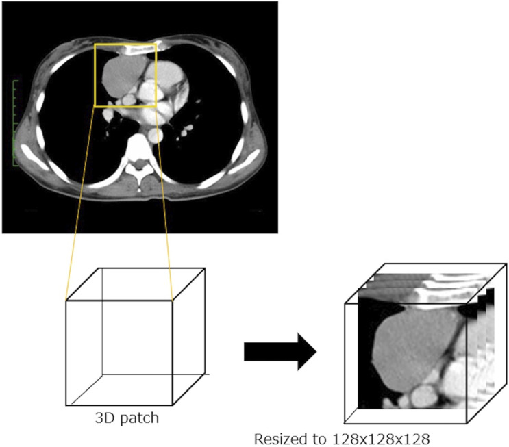
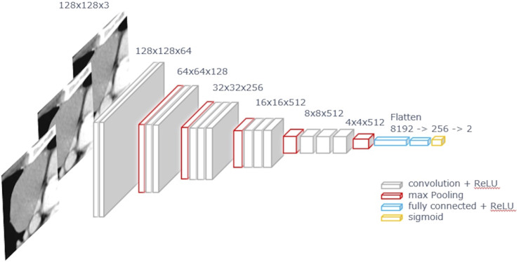
159 patients with 160 thymomas were included. A fine-tuning VGG16 network model with Adam optimizer was used, followed by k-fold cross validation. The dataset consisted of three axial slices, including the maximum tumor size from the CT volume data. The data were augmented 50 times by rotation, zoom, shear, and horizontal/vertical flip. Three independent networks for the CT dataset were considered, and the result was determined by voting. Three radiologists independently diagnosed thymomas with and without the model. The area under the curve (AUC) of the diagnostic performance was compared using receiver operating characteristic analysis.

RESULTS

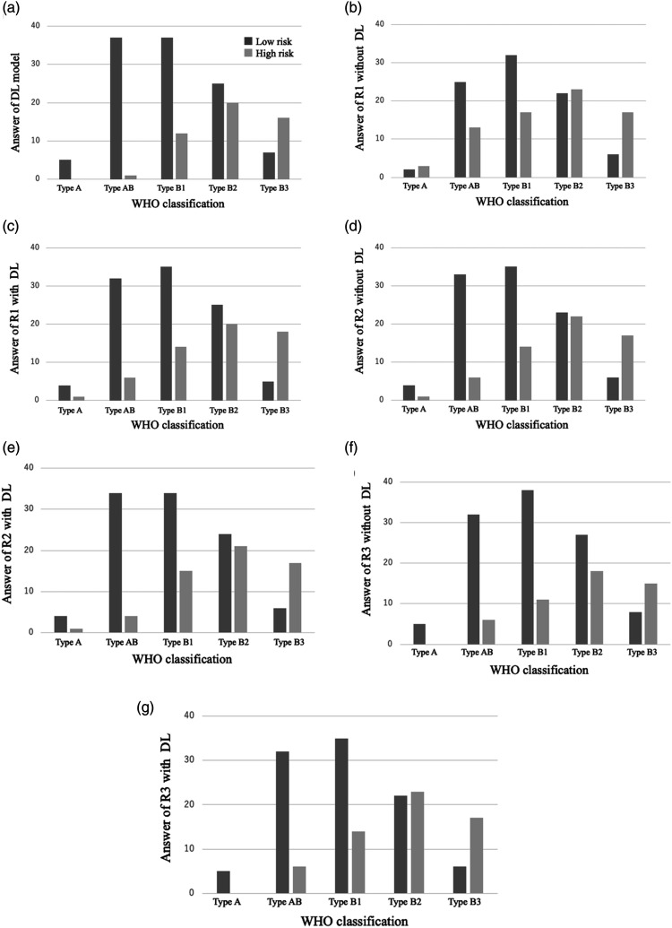
Accuracy of the DL model was 71.3%. Diagnostic performance of the radiologists was as follows: AUC and accuracy without the DL model, 0.61-0.68 and 61.9%-69.3%; and with the DL model, 0.66-0.69 and 68.1%-70.0%, respectively. AUC of the diagnostic performance showed no significant differences between radiologists with and without the DL model. The DL model tended to increase the diagnostic accuracy, but AUC was not significantly improved.

CONCLUSION

Diagnostic performance of the DL was comparable to that of radiologists. The DL model assistance tended to increase diagnostic accuracy.

摘要

背景

根据世界卫生组织的分类,基于CT的深度学习(DL)对胸腺瘤的研究较少。

目的

开发一种基于CT的DL模型,以区分低风险和高风险胸腺瘤,并比较使用和不使用DL模型时放射科医生的诊断性能。

材料与方法

纳入159例患者的160个胸腺瘤。使用带有Adam优化器的微调VGG16网络模型,随后进行k折交叉验证。数据集由三个轴向切片组成,包括来自CT体积数据的最大肿瘤大小。数据通过旋转、缩放、剪切和水平/垂直翻转进行50次增强。考虑了用于CT数据集的三个独立网络,并通过投票确定结果。三位放射科医生分别在有和没有该模型的情况下独立诊断胸腺瘤。使用受试者操作特征分析比较诊断性能的曲线下面积(AUC)。

结果

DL模型的准确率为71.3%。放射科医生的诊断性能如下:不使用DL模型时,AUC和准确率分别为0.61 - 0.68和61.9% - 69.3%;使用DL模型时,分别为0.66 - 0.69和68.1% - 70.0%。有和没有DL模型的放射科医生之间,诊断性能的AUC没有显著差异。DL模型倾向于提高诊断准确性,但AUC没有显著改善。

结论

DL的诊断性能与放射科医生相当。DL模型辅助倾向于提高诊断准确性。

https://cdn.ncbi.nlm.nih.gov/pmc/blobs/586a/11457241/82575f3ccb65/10.1177_20584601241288509-fig4.jpg
https://cdn.ncbi.nlm.nih.gov/pmc/blobs/586a/11457241/62fdd6e3d1d0/10.1177_20584601241288509-fig1.jpg
https://cdn.ncbi.nlm.nih.gov/pmc/blobs/586a/11457241/d9ddb4d5acb0/10.1177_20584601241288509-fig2.jpg
https://cdn.ncbi.nlm.nih.gov/pmc/blobs/586a/11457241/09e42a08c41c/10.1177_20584601241288509-fig3.jpg
https://cdn.ncbi.nlm.nih.gov/pmc/blobs/586a/11457241/82575f3ccb65/10.1177_20584601241288509-fig4.jpg

相似文献

1
Differential diagnosis between low-risk and high-risk thymoma: Comparison of diagnostic performance of radiologists with and without deep learning model.低风险与高风险胸腺瘤的鉴别诊断:有深度学习模型和无深度学习模型的放射科医生诊断性能比较
Acta Radiol Open. 2024 Oct 4;13(10):20584601241288509. doi: 10.1177/20584601241288509. eCollection 2024 Oct.
2
Development of a deep learning model for classifying thymoma as Masaoka-Koga stage I or II via preoperative CT images.通过术前CT图像开发用于将胸腺瘤分类为Masaoka-Koga I期或II期的深度学习模型。
Ann Transl Med. 2020 Mar;8(6):287. doi: 10.21037/atm.2020.02.183.
3
Differentiating adrenal metastases from benign lesions with multiphase CT imaging: Deep learning could play an active role in assisting radiologists.多期 CT 成像鉴别肾上腺转移瘤与良性病变:深度学习在协助放射科医生方面可能发挥积极作用。
Eur J Radiol. 2023 Dec;169:111169. doi: 10.1016/j.ejrad.2023.111169. Epub 2023 Nov 7.
4
Deep Learning in Prostate Cancer Diagnosis Using Multiparametric Magnetic Resonance Imaging With Whole-Mount Histopathology Referenced Delineations.基于全层组织病理学参考勾画的多参数磁共振成像在前列腺癌诊断中的深度学习
Front Med (Lausanne). 2022 Jan 13;8:810995. doi: 10.3389/fmed.2021.810995. eCollection 2021.
5
Development and Validation of Contrast-Enhanced CT-Based Deep Transfer Learning and Combined Clinical-Radiomics Model to Discriminate Thymomas and Thymic Cysts: A Multicenter Study.基于增强 CT 的深度迁移学习与联合临床放射组学模型在鉴别胸腺瘤和胸腺囊肿中的建立与验证:一项多中心研究。
Acad Radiol. 2024 Apr;31(4):1615-1628. doi: 10.1016/j.acra.2023.10.018. Epub 2023 Nov 10.
6
Deep convolutional neural network for classification of thyroid nodules on ultrasound: Comparison of the diagnostic performance with that of radiologists.基于超声的甲状腺结节深度学习卷积神经网络分类:与放射科医生诊断效能比较
Eur J Radiol. 2022 Jul;152:110335. doi: 10.1016/j.ejrad.2022.110335. Epub 2022 Apr 30.
7
Deep learning for the diagnosis of suspicious thyroid nodules based on multimodal ultrasound images.基于多模态超声图像的深度学习用于可疑甲状腺结节的诊断
Front Oncol. 2022 Nov 8;12:1012724. doi: 10.3389/fonc.2022.1012724. eCollection 2022.
8
Radiomics Signatures of Computed Tomography Imaging for Predicting Risk Categorization and Clinical Stage of Thymomas.基于 CT 影像学的影像组学特征预测胸腺瘤风险分级和临床分期
Biomed Res Int. 2019 May 28;2019:3616852. doi: 10.1155/2019/3616852. eCollection 2019.
9
Deep learning-assisted diagnosis of benign and malignant parotid tumors based on ultrasound: a retrospective study.基于超声的深度学习辅助诊断腮腺良恶性肿瘤:一项回顾性研究。
BMC Cancer. 2024 Apr 23;24(1):510. doi: 10.1186/s12885-024-12277-8.
10
CT-based deep learning model to differentiate invasive pulmonary adenocarcinomas appearing as subsolid nodules among surgical candidates: comparison of the diagnostic performance with a size-based logistic model and radiologists.基于 CT 的深度学习模型用于区分可手术患者中表现为亚实性结节的浸润性肺腺癌:与基于大小的逻辑模型和放射科医生相比,该模型的诊断性能比较。
Eur Radiol. 2020 Jun;30(6):3295-3305. doi: 10.1007/s00330-019-06628-4. Epub 2020 Feb 13.

本文引用的文献

1
Dual-energy CT perfusion imaging for differentiating WHO subtypes of thymic epithelial tumors.双能量 CT 灌注成像在胸腺瘤 WHO 组织学分型中的鉴别诊断价值。
Sci Rep. 2020 Mar 26;10(1):5511. doi: 10.1038/s41598-020-62466-1.
2
Deep learning: definition and perspectives for thoracic imaging.深度学习:胸部影像学的定义和展望。
Eur Radiol. 2020 Apr;30(4):2021-2030. doi: 10.1007/s00330-019-06564-3. Epub 2019 Dec 6.
3
Quantitative 3D Shape Analysis of CT Images of Thymoma: A Comparison With Histological Types.胸腺瘤 CT 图像的定量 3D 形态分析:与组织学分型的比较。
AJR Am J Roentgenol. 2020 Feb;214(2):341-347. doi: 10.2214/AJR.19.21844. Epub 2019 Nov 6.
4
Predicting EGFR mutation status in lung adenocarcinoma on computed tomography image using deep learning.利用深度学习技术在 CT 图像上预测肺腺癌中的 EGFR 突变状态。
Eur Respir J. 2019 Mar 28;53(3). doi: 10.1183/13993003.00986-2018. Print 2019 Mar.
5
Deep learning for classifying fibrotic lung disease on high-resolution computed tomography: a case-cohort study.深度学习在高分辨率 CT 上对肺纤维化疾病的分类:一项病例队列研究。
Lancet Respir Med. 2018 Nov;6(11):837-845. doi: 10.1016/S2213-2600(18)30286-8. Epub 2018 Sep 16.
6
Thymic epithelial tumours: ESMO Clinical Practice Guidelines for diagnosis, treatment and follow-up.胸腺上皮肿瘤:ESMO诊断、治疗及随访临床实践指南
Ann Oncol. 2015 Sep;26 Suppl 5:v40-55. doi: 10.1093/annonc/mdv277.
7
Associations between computed tomography features of thymomas and their pathological classification.胸腺瘤的计算机断层扫描特征与其病理分类之间的关联。
Acta Radiol. 2016 Nov;57(11):1318-1325. doi: 10.1177/0284185115590288. Epub 2016 Jul 20.
8
Predicting subtypes of thymic epithelial tumors using CT: new perspective based on a comprehensive analysis of 216 patients.利用CT预测胸腺瘤上皮肿瘤亚型:基于对216例患者的综合分析的新视角
Sci Rep. 2014 Nov 10;4:6984. doi: 10.1038/srep06984.
9
ITMIG consensus statement on the use of the WHO histological classification of thymoma and thymic carcinoma: refined definitions, histological criteria, and reporting.国际胸腺肿瘤学会共识声明:关于采用世界卫生组织胸腺瘤和胸腺癌组织学分类的使用:精细定义、组织学标准和报告。
J Thorac Oncol. 2014 May;9(5):596-611. doi: 10.1097/JTO.0000000000000154.
10
Relationship between computed tomography manifestations of thymic epithelial tumors and the WHO pathological classification.胸腺上皮肿瘤的计算机断层扫描表现与世界卫生组织病理分类之间的关系
Asian Pac J Cancer Prev. 2012;13(11):5581-5. doi: 10.7314/apjcp.2012.13.11.5581.