• 文献检索
  • 文档翻译
  • 深度研究
  • 学术资讯
  • Suppr Zotero 插件Zotero 插件
  • 邀请有礼
  • 套餐&价格
  • 历史记录
应用&插件
Suppr Zotero 插件Zotero 插件浏览器插件Mac 客户端Windows 客户端微信小程序
定价
高级版会员购买积分包购买API积分包
服务
文献检索文档翻译深度研究API 文档MCP 服务
关于我们
关于 Suppr公司介绍联系我们用户协议隐私条款
关注我们

Suppr 超能文献

核心技术专利:CN118964589B侵权必究
粤ICP备2023148730 号-1Suppr @ 2026

文献检索

告别复杂PubMed语法,用中文像聊天一样搜索,搜遍4000万医学文献。AI智能推荐,让科研检索更轻松。

立即免费搜索

文件翻译

保留排版,准确专业,支持PDF/Word/PPT等文件格式,支持 12+语言互译。

免费翻译文档

深度研究

AI帮你快速写综述,25分钟生成高质量综述,智能提取关键信息,辅助科研写作。

立即免费体验

多发性硬化症中顺磁性边缘病变异质性的临床相关性

Clinical relevance of paramagnetic rim lesion heterogeneity in multiple sclerosis.

作者信息

Stölting Anna, Vanden Bulcke Colin, Borrelli Serena, Bugli Céline, Du Pasquier Renaud, van Pesch Vincent, Maggi Pietro

机构信息

Neuroinflammation Imaging Lab (NIL), Institute of NeuroScience, Université catholique de Louvain, Brussels, Belgium.

ICTEAM Institute, Université catholique de Louvain, Louvain-la-Neuve, Belgium.

出版信息

Ann Clin Transl Neurol. 2024 Dec;11(12):3137-3151. doi: 10.1002/acn3.52220. Epub 2024 Oct 9.

DOI:10.1002/acn3.52220
PMID:39382072
原文链接:https://pmc.ncbi.nlm.nih.gov/articles/PMC11651185/
Abstract

OBJECTIVE

Previous studies reveal heterogeneity in terms of paramagnetic rim lesions (PRL) associated tissue damage. We investigated the physiopathology and clinical implications of this heterogeneity.

METHODS

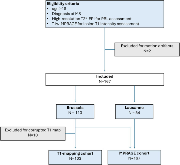
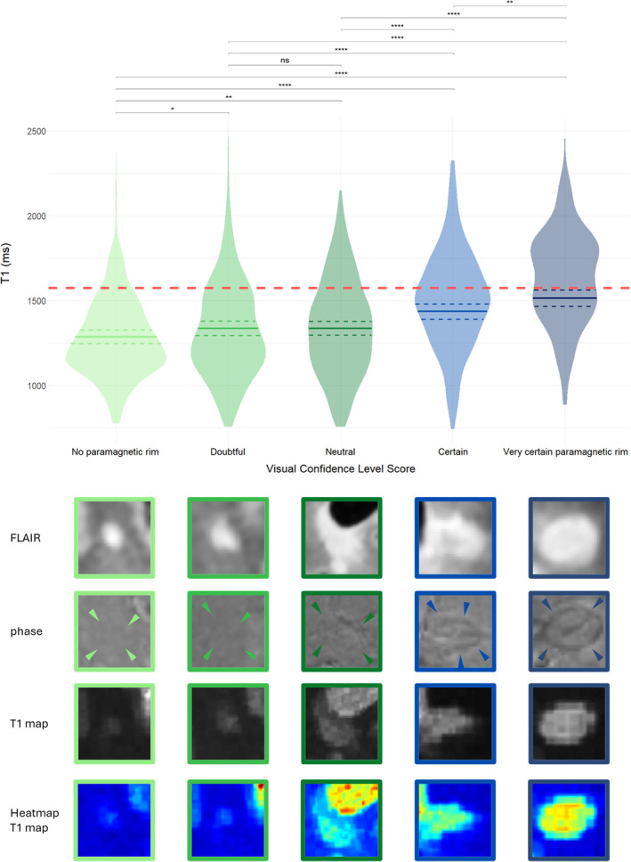
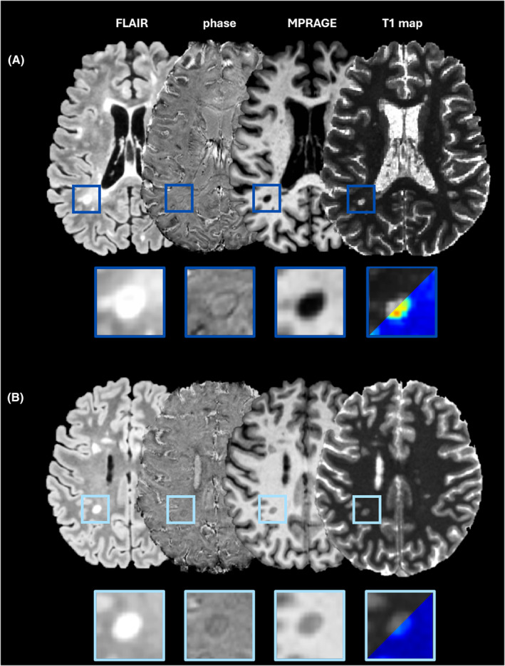
In 103 MS patients (72 relapsing and 31 progressive), brain lesions were manually segmented on 3T 3D-FLAIR and rim visibility was assessed with a visual confidence level score (VCLS) on 3D-EPI phase. Using T1 relaxation time maps, lesions were categorized in long-T1 and short-T1. Lesion age was calculated from time of first gadolinium enhancement (N = 84 lesions). Results on clinical scores were validated in an extended cohort of 167 patients using normalized T1w-MPRAGE lesion values.

RESULTS

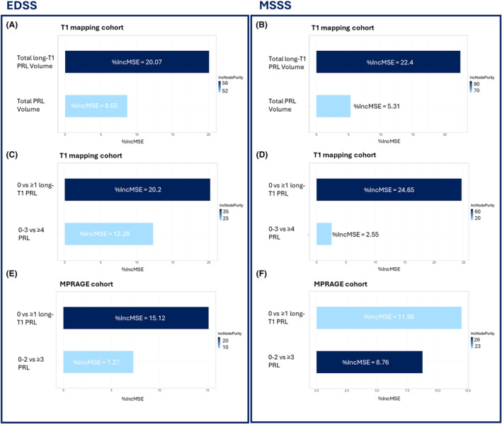
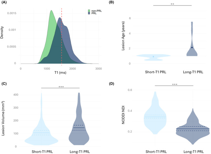
Rim visibility (VCLS analysis) was associated with increasing lesional T1 (P/P < 0.001). Of 1680 analyzed lesions, 427 were categorized as PRL. Long-T1 PRL were older than short-T1 PRL (average 0.8 vs. 2.0 years, P/P = 0.005/0.008), and featured larger lesional volume (P/P < 0.0001) and multi-shell diffusion-measured axonal damage (P/P < 0.0001). The total volume of long-T1-PRL versus PRL showed 2× predictive power for both higher MS disability (EDSS; P/P = 0.003/0.005 vs. P/P = 0.042/0.057) and severity (MSSS; P/P = 0.0006/0.001 vs. P/P = 0.004/0.007). In random forest, having ≥1 long-T1-PRL versus ≥4 PRL showed 2-4× higher performance to predict a higher EDSS and MSSS. In the validation cohort, long-T1 PRL outperformed (~2×) PRL in predicting both EDSS and MSSS.

INTERPRETATION

PRL show substantial heterogeneity in terms of intralesional tissue damage. More destructive, likely older, long-T1 PRL improve the association with MS clinical scales. This PRL heterogeneity characterization was replicated using standard T1w MRI, highlighting its potential for clinical translation.

摘要

目的

既往研究揭示了与顺磁性边缘病变(PRL)相关的组织损伤存在异质性。我们调查了这种异质性的生理病理学及临床意义。

方法

对103例多发性硬化症(MS)患者(72例复发型和31例进展型),在3T 3D-FLAIR序列上手动分割脑病变,并在3D-EPI相位上用视觉置信度评分(VCLS)评估边缘可见性。利用T1弛豫时间图,将病变分为长T1和短T1两类。病变年龄根据首次钆增强时间计算(N = 84个病变)。使用标准化的T1w-MPRAGE病变值,在167例患者的扩展队列中验证临床评分结果。

结果

边缘可见性(VCLS分析)与病变T1增加相关(P/P < 0.001)。在1680个分析的病变中,427个被归类为PRL。长T1 PRL比短T1 PRL年龄更大(平均0.8年对2.0年,P/P = 0.005/0.008),且具有更大的病变体积(P/P < 0.0001)和多壳扩散测量的轴突损伤(P/P < 0.0001)。长T1-PRL与PRL的总体积对更高的MS残疾(扩展残疾状态量表;EDSS;P/P = 0.003/0.005对P/P = 0.042/0.057)和严重程度(多发性硬化症严重程度量表;MSSS;P/P = 0.0006/0.001对P/P = 0.004/0.007)均显示出2倍的预测能力。在随机森林分析中,有≥1个长T1-PRL与≥4个PRL相比,在预测更高的EDSS和MSSS方面表现出高2 - 4倍的性能。在验证队列中,长T1 PRL在预测EDSS和MSSS方面优于PRL(约2倍)。

解读

PRL在病变内组织损伤方面表现出显著的异质性。更具破坏性、可能更陈旧的长T1 PRL改善了与MS临床量表的相关性。这种PRL异质性特征通过标准T1w MRI得以复制,突出了其临床转化的潜力。

https://cdn.ncbi.nlm.nih.gov/pmc/blobs/8bba/11651185/ceff3eb4ce90/ACN3-11-3137-g001.jpg
https://cdn.ncbi.nlm.nih.gov/pmc/blobs/8bba/11651185/92c0f1ee3ef0/ACN3-11-3137-g004.jpg
https://cdn.ncbi.nlm.nih.gov/pmc/blobs/8bba/11651185/f9e1ed5e5b31/ACN3-11-3137-g002.jpg
https://cdn.ncbi.nlm.nih.gov/pmc/blobs/8bba/11651185/14c3e8dd6de7/ACN3-11-3137-g005.jpg
https://cdn.ncbi.nlm.nih.gov/pmc/blobs/8bba/11651185/76a51c6cb22d/ACN3-11-3137-g003.jpg
https://cdn.ncbi.nlm.nih.gov/pmc/blobs/8bba/11651185/ceff3eb4ce90/ACN3-11-3137-g001.jpg

相似文献

1
Clinical relevance of paramagnetic rim lesion heterogeneity in multiple sclerosis.多发性硬化症中顺磁性边缘病变异质性的临床相关性
Ann Clin Transl Neurol. 2024 Dec;11(12):3137-3151. doi: 10.1002/acn3.52220. Epub 2024 Oct 9.
2
Characterization of white matter lesions in multiple sclerosis using proton density and T1-relaxation measures.使用质子密度和 T1 弛豫测量对多发性硬化症的脑白质病变进行特征描述。
Magn Reson Imaging. 2024 Feb;106:110-118. doi: 10.1016/j.mri.2023.12.004. Epub 2023 Dec 23.
3
Comparative overview of multi-shell diffusion MRI models to characterize the microstructure of multiple sclerosis lesions and periplaques.多壳扩散 MRI 模型对多发性硬化病变及斑块微结构特征的比较综述。
Neuroimage Clin. 2024;42:103593. doi: 10.1016/j.nicl.2024.103593. Epub 2024 Mar 18.
4
Long-term evolution of multiple sclerosis iron rim lesions in 7 T MRI.7T MRI 下多发性硬化症铁环病变的长期演变。
Brain. 2021 Apr 12;144(3):833-847. doi: 10.1093/brain/awaa436.
5
T1- Thresholds in Black Holes Increase Clinical-Radiological Correlation in Multiple Sclerosis Patients.T1 - 黑洞中的阈值增加了多发性硬化症患者的临床 - 放射学相关性。
PLoS One. 2015 Dec 11;10(12):e0144693. doi: 10.1371/journal.pone.0144693. eCollection 2015.
6
Spinal cord lesions and brain grey matter atrophy independently predict clinical worsening in definite multiple sclerosis: a 5-year, multicentre study.脊髓病变和脑灰质萎缩独立预测明确多发性硬化症的临床恶化:一项为期5年的多中心研究。
J Neurol Neurosurg Psychiatry. 2023 Jan;94(1):10-18. doi: 10.1136/jnnp-2022-329854. Epub 2022 Sep 28.
7
Personalized maps of T1 relaxometry abnormalities provide correlates of disability in multiple sclerosis patients.个性化 T1 弛豫率异常图谱为多发性硬化症患者的残疾提供相关信息。
Neuroimage Clin. 2023;37:103349. doi: 10.1016/j.nicl.2023.103349. Epub 2023 Feb 13.
8
Deeply 3D-T1-TFE hypointense voxels are characteristic of phase-rim lesions in multiple sclerosis.深度三维 T1-TFE 低信号体素是多发性硬化症相位边缘病变的特征。
Eur Radiol. 2024 Feb;34(2):1337-1345. doi: 10.1007/s00330-023-09784-w. Epub 2023 Jun 6.
9
Slowly expanding lesions relate to persisting black-holes and clinical outcomes in relapse-onset multiple sclerosis.在复发缓解型多发性硬化症中,缓慢扩大的病灶与持续存在的黑洞和临床结局有关。
Neuroimage Clin. 2022;35:103048. doi: 10.1016/j.nicl.2022.103048. Epub 2022 May 16.
10
Neurological disability and brain grey matter atrophy in primary progressive multiple sclerosis are determined by microstructural lesional changes, but not by lesion load.原发性进行性多发性硬化症中的神经功能障碍和脑灰质萎缩是由微观结构病变改变决定的,而非由病灶负荷决定。
J Neurol. 2025 Apr 1;272(4):302. doi: 10.1007/s00415-025-13043-x.

引用本文的文献

1
Paramagnetic rim lesions are highly specific for multiple sclerosis in real-world data.在实际数据中,顺磁性边缘病变对多发性硬化具有高度特异性。
Brain Commun. 2025 May 29;7(3):fcaf211. doi: 10.1093/braincomms/fcaf211. eCollection 2025.

本文引用的文献

1
Central Vein Sign, Cortical Lesions, and Paramagnetic Rim Lesions for the Diagnostic and Prognostic Workup of Multiple Sclerosis.中央静脉征、皮质病变和顺磁性边缘病变在多发性硬化的诊断和预后评估中的作用。
Neurol Neuroimmunol Neuroinflamm. 2024 Jul;11(4):e200253. doi: 10.1212/NXI.0000000000200253. Epub 2024 May 24.
2
Comparative overview of multi-shell diffusion MRI models to characterize the microstructure of multiple sclerosis lesions and periplaques.多壳扩散 MRI 模型对多发性硬化病变及斑块微结构特征的比较综述。
Neuroimage Clin. 2024;42:103593. doi: 10.1016/j.nicl.2024.103593. Epub 2024 Mar 18.
3
Imaging chronic active lesions in multiple sclerosis: a consensus statement.
多发性硬化症慢性活动性病变的影像学:共识声明。
Brain. 2024 Sep 3;147(9):2913-2933. doi: 10.1093/brain/awae013.
4
Association of Spinal Cord Atrophy and Brain Paramagnetic Rim Lesions With Progression Independent of Relapse Activity in People With MS.脊髓萎缩和脑磁共振边缘信号异常与 MS 患者的疾病进展相关,与复发活动无关。
Neurology. 2024 Jan 9;102(1):e207768. doi: 10.1212/WNL.0000000000207768. Epub 2023 Dec 13.
5
Harmonizing Definitions for Progression Independent of Relapse Activity in Multiple Sclerosis: A Systematic Review.统一多发性硬化症中与复发活动无关的进展的定义:一项系统评价
JAMA Neurol. 2023 Nov 1;80(11):1232-1245. doi: 10.1001/jamaneurol.2023.3331.
6
Paramagnetic rim lesions lead to pronounced diffuse periplaque white matter damage in multiple sclerosis.顺磁边缘病变导致多发性硬化症斑块周围白质弥漫性损伤明显。
Mult Scler. 2023 Oct;29(11-12):1406-1417. doi: 10.1177/13524585231197954. Epub 2023 Sep 15.
7
B cell depletion therapy does not resolve chronic active multiple sclerosis lesions.B 细胞耗竭疗法不能解决慢性活动性多发性硬化症病灶。
EBioMedicine. 2023 Aug;94:104701. doi: 10.1016/j.ebiom.2023.104701. Epub 2023 Jul 10.
8
A New Advanced MRI Biomarker for Remyelinated Lesions in Multiple Sclerosis.一种用于多发性硬化症中髓鞘再生病变的新型高级 MRI 生物标志物。
Ann Neurol. 2022 Sep;92(3):486-502. doi: 10.1002/ana.26441. Epub 2022 Jul 13.
9
Long-term dynamics of multiple sclerosis iron rim lesions.多发性硬化症铁环病变的长期动态变化。
Mult Scler Relat Disord. 2022 Jan;57:103340. doi: 10.1016/j.msard.2021.103340. Epub 2021 Oct 18.
10
Smouldering multiple sclerosis: the 'real MS'.隐匿性多发性硬化症:“真正的多发性硬化症”
Ther Adv Neurol Disord. 2022 Jan 25;15:17562864211066751. doi: 10.1177/17562864211066751. eCollection 2022.