Oxford Eye Hospital, John Radcliffe Hospital, Oxford University Hospitals NHS Foundation Trust, Oxford, OX3 9DU, UK.
Nuffield Laboratory of Ophthalmology, Nuffield Department of Clinical Neurosciences, Level 6 John Radcliffe Hospital, University of Oxford, Headley Way, Oxford, OX3 9DU, UK.
Sci Rep. 2024 Oct 9;14(1):23629. doi: 10.1038/s41598-024-74088-y.
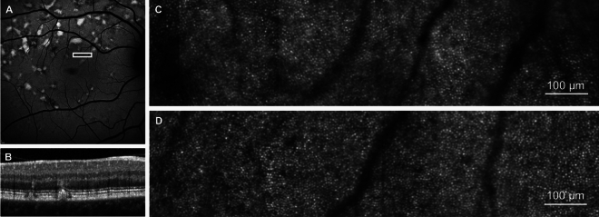
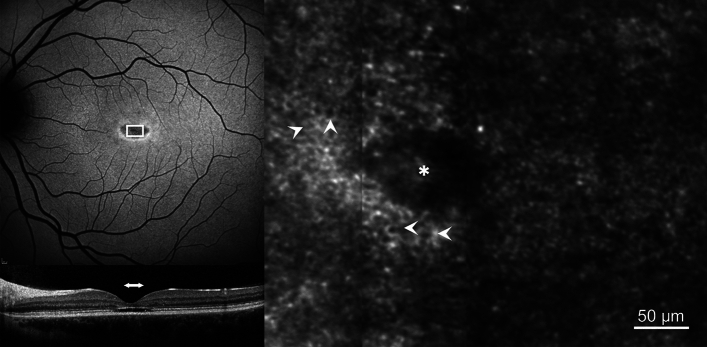
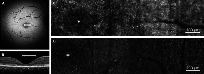
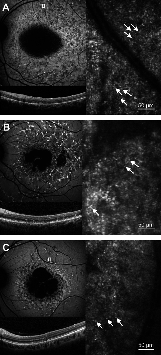
Image based cell-specific biomarkers will play an important role in monitoring treatment outcomes of novel therapies in patients with Stargardt (STGD1) disease and may provide information on the exact mechanism of retinal degeneration. This study reports retinal image features from conventional clinical imaging and from corresponding high-resolution imaging with a confocal adaptive optics scanning laser ophthalmoscope (AOSLO) in a heterogenous cohort of patients with Stargardt (STGD1) disease. This is a prospective observational study in which 16 participants with clinically and molecularly confirmed STGD1, and 7 healthy controls underwent clinical assessment and confocal AOSLO imaging. Clinical assessment included short-wavelength and near-infrared fundus autofluorescence, spectral-domain optical coherence tomography, and macular microperimetry. AOSLO images were acquired over a range of retinal eccentricities (0°-20°) and mapped to areas of interest from the clinical images. A regular photoreceptor mosaic was identified in areas of normal or near normal retinal structure on clinical images. Where clinical imaging indicated areas of retinal degeneration, the photoreceptor mosaic was disorganised and lacked unambiguous cones. Discrete hyper-reflective foci were identified in 9 participants with STGD1 within areas of retinal degeneration. A continuous RPE cell mosaic at the fovea was identified in one participant with an optical gap phenotype. The clinical heterogeneity observed in STGD1 is reflected in the findings on confocal AOSLO imaging.
基于图像的细胞特异性生物标志物将在监测新型疗法治疗斯塔加德特(STGD1)病患者的治疗效果方面发挥重要作用,并可能提供有关视网膜变性的确切机制的信息。本研究报告了来自常规临床成像和共焦自适应光学扫描激光检眼镜(AOSLO)的对应高分辨率成像的视网膜图像特征,该研究涉及异质队列的斯塔加德特(STGD1)病患者。这是一项前瞻性观察性研究,其中 16 名经临床和分子证实的 STGD1 患者和 7 名健康对照者接受了临床评估和共焦 AOSLO 成像。临床评估包括短波和近红外眼底自发荧光、谱域光学相干断层扫描和黄斑微视野计。AOSLO 图像在一系列视网膜偏心度(0°-20°)上获取,并与临床图像的感兴趣区域映射。在临床图像上显示正常或接近正常视网膜结构的区域中,识别出规则的光感受器马赛克。在临床成像指示视网膜变性的区域中,光感受器马赛克紊乱且缺乏明确的视锥细胞。在 9 名 STGD1 患者的视网膜变性区域中发现了离散的高反射焦点。在具有光学间隙表型的一名参与者中,在黄斑区识别到连续的 RPE 细胞马赛克。在 STGD1 中观察到的临床异质性反映在共焦 AOSLO 成像的结果中。