• 文献检索
  • 文档翻译
  • 深度研究
  • 学术资讯
  • Suppr Zotero 插件Zotero 插件
  • 邀请有礼
  • 套餐&价格
  • 历史记录
应用&插件
Suppr Zotero 插件Zotero 插件浏览器插件Mac 客户端Windows 客户端微信小程序
定价
高级版会员购买积分包购买API积分包
服务
文献检索文档翻译深度研究API 文档MCP 服务
关于我们
关于 Suppr公司介绍联系我们用户协议隐私条款
关注我们

Suppr 超能文献

核心技术专利:CN118964589B侵权必究
粤ICP备2023148730 号-1Suppr @ 2026

文献检索

告别复杂PubMed语法,用中文像聊天一样搜索,搜遍4000万医学文献。AI智能推荐,让科研检索更轻松。

立即免费搜索

文件翻译

保留排版,准确专业,支持PDF/Word/PPT等文件格式,支持 12+语言互译。

免费翻译文档

深度研究

AI帮你快速写综述,25分钟生成高质量综述,智能提取关键信息,辅助科研写作。

立即免费体验

使用吉布斯振铃衰减的T1加权/ T2加权液体衰减反转恢复序列髓鞘MRI绘制皮质层特定区域边界

Charting cortical-layer specific area boundaries using Gibbs' ringing attenuated T1w/T2w-FLAIR myelin MRI.

作者信息

Autio Joonas A, Uematsu Akiko, Ikeda Takuro, Ose Takayuki, Hou Yujie, Magrou Loïc, Kimura Ikko, Ohno Masahiro, Murata Katsutoshi, Coalson Tim, Kennedy Henry, Glasser Matthew F, Van Essen David C, Hayashi Takuya

机构信息

Laboratory for Brain Connectomics Imaging, RIKEN Center for Biosystems Dynamics Research, Kobe, Japan.

Université Lyon, Université Claude Bernard Lyon 1, Inserm, Stem Cell and Brain Research Institute U1208, 69500, Bron, France.

出版信息

bioRxiv. 2024 Nov 13:2024.09.27.615294. doi: 10.1101/2024.09.27.615294.

DOI:10.1101/2024.09.27.615294
PMID:39386722
原文链接:https://pmc.ncbi.nlm.nih.gov/articles/PMC11463467/
Abstract

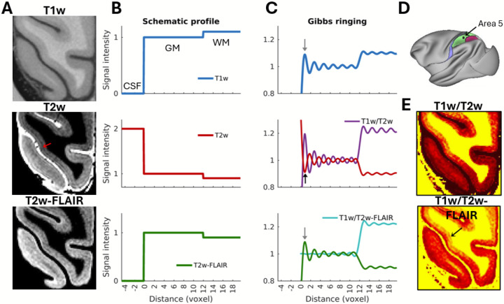
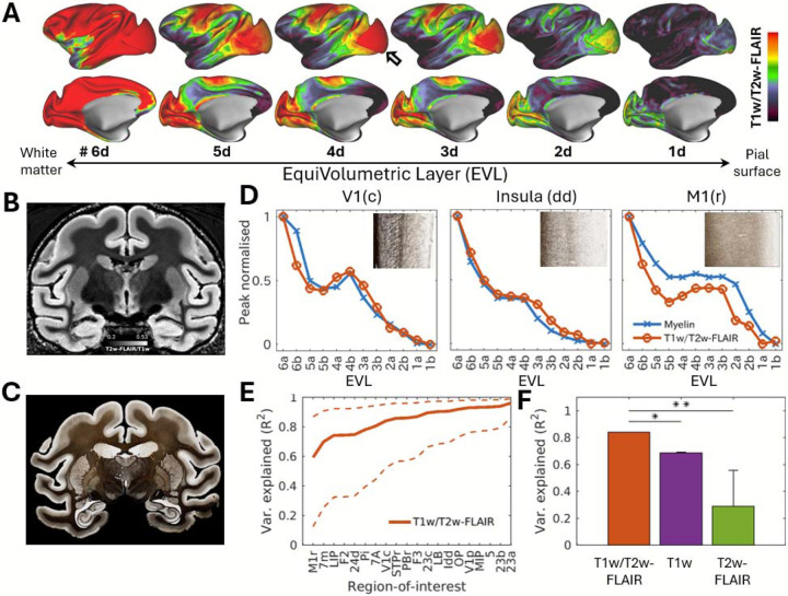
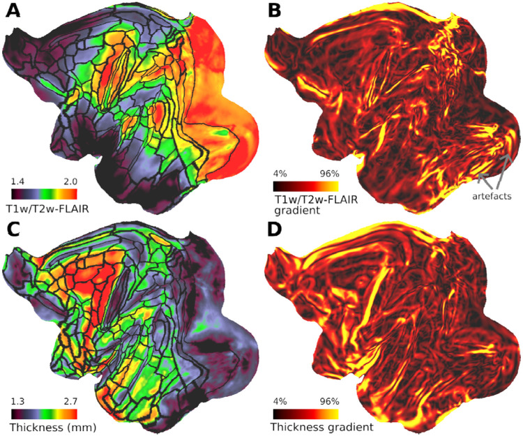
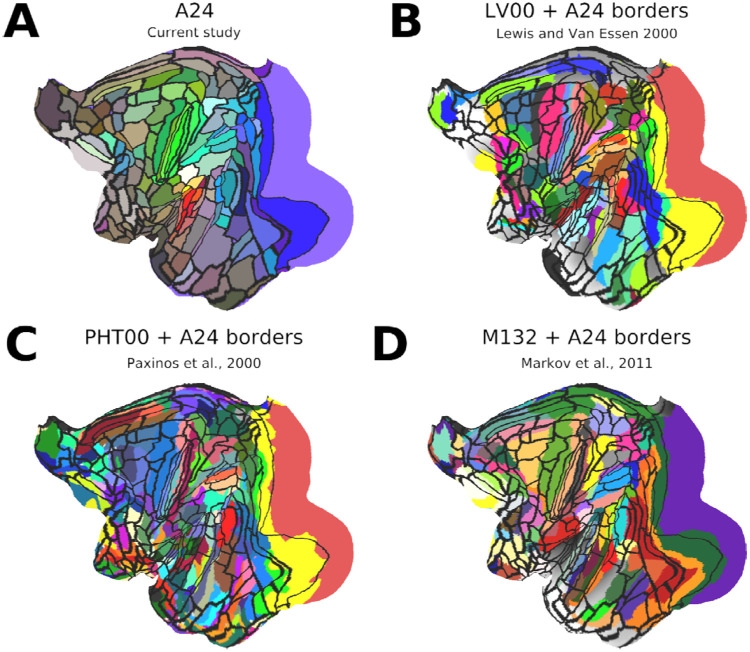
Cortical areas have traditionally been defined by their distinctive layer cyto- and/or myelo- architecture using postmortem histology. Recent studies have delineated many areas by measuring overall cortical myelin content and its spatial gradients using the T1w/T2w ratio MRI in living primates, including humans. While T1w/T2w studies of areal transitions might benefit from using the layer profile of this myelin-related contrast, a significant confound is Gibbs' ringing artefact, which produces signal fluctuations resembling cortical layers. Here, we address these issues with a novel approach using cortical layer thickness-adjusted T1w/T2w-FLAIR imaging, which effectively cancels out Gibbs' ringing artefacts while enhancing intra-cortical myelin contrast. Whole-brain MRI measures were mapped onto twelve equivolumetric layers, and layer-specific sharp myeloarchitectonic transitions were identified using spatial gradients resulting in a putative 182 area/subarea partition of the macaque cerebral cortex. The myelin maps exhibit notably high homology with those in humans, suggesting cortical myelin shares a similar developmental program across species. Comparison with histological Gallyas myelin stains explains over 80% of the variance in the laminar T1w/T2w-FLAIR profiles, substantiating the validity of the method. Altogether, our approach provides a novel, noninvasive means for precision mapping layer myeloarchitecture in the primate cerebral cortex, advancing the pioneering work of classical neuroanatomists.

摘要

传统上,皮质区域是通过死后组织学,依据其独特的层状细胞结构和/或髓鞘结构来定义的。最近的研究通过在包括人类在内的活体灵长类动物中使用T1w/T2w比率MRI测量整体皮质髓鞘含量及其空间梯度,描绘出了许多区域。虽然关于区域过渡的T1w/T2w研究可能会受益于使用这种与髓鞘相关对比度的层状轮廓,但一个重大的混淆因素是吉布斯振铃伪影,它会产生类似于皮质层的信号波动。在这里,我们采用一种新颖的方法,即使用皮质层厚度调整后的T1w/T2w-FLAIR成像来解决这些问题,这种方法在增强皮质内髓鞘对比度的同时,有效地消除了吉布斯振铃伪影。全脑MRI测量结果被映射到十二个等体积层上,并使用空间梯度识别出特定层的尖锐髓鞘结构过渡,从而得出猕猴大脑皮质的一个假定的182个区域/子区域划分。髓鞘图谱与人类的图谱显示出显著的高度同源性,表明皮质髓鞘在不同物种间共享相似的发育程序。与组织学Gallyas髓鞘染色的比较解释了层状T1w/T2w-FLAIR图谱中超过80%的差异,证实了该方法的有效性。总之,我们的方法为精确绘制灵长类动物大脑皮质的层状髓鞘结构提供了一种新颖的非侵入性手段,推进了经典神经解剖学家的开创性工作。

https://cdn.ncbi.nlm.nih.gov/pmc/blobs/2228/11562190/60f367eb4d18/nihpp-2024.09.27.615294v3-f0007.jpg
https://cdn.ncbi.nlm.nih.gov/pmc/blobs/2228/11562190/9b37005a2c6d/nihpp-2024.09.27.615294v3-f0001.jpg
https://cdn.ncbi.nlm.nih.gov/pmc/blobs/2228/11562190/ed9c5fa39e32/nihpp-2024.09.27.615294v3-f0002.jpg
https://cdn.ncbi.nlm.nih.gov/pmc/blobs/2228/11562190/8c3544d49864/nihpp-2024.09.27.615294v3-f0003.jpg
https://cdn.ncbi.nlm.nih.gov/pmc/blobs/2228/11562190/db86be32648c/nihpp-2024.09.27.615294v3-f0004.jpg
https://cdn.ncbi.nlm.nih.gov/pmc/blobs/2228/11562190/09d2ed9d0382/nihpp-2024.09.27.615294v3-f0005.jpg
https://cdn.ncbi.nlm.nih.gov/pmc/blobs/2228/11562190/5720443e60a8/nihpp-2024.09.27.615294v3-f0006.jpg
https://cdn.ncbi.nlm.nih.gov/pmc/blobs/2228/11562190/60f367eb4d18/nihpp-2024.09.27.615294v3-f0007.jpg

相似文献

1
Charting cortical-layer specific area boundaries using Gibbs' ringing attenuated T1w/T2w-FLAIR myelin MRI.使用吉布斯振铃衰减的T1加权/ T2加权液体衰减反转恢复序列髓鞘MRI绘制皮质层特定区域边界
bioRxiv. 2024 Nov 13:2024.09.27.615294. doi: 10.1101/2024.09.27.615294.
2
Gray matter myelination of 1555 human brains using partial volume corrected MRI images.使用部分容积校正MRI图像对1555个人脑进行灰质髓鞘化研究。
Neuroimage. 2015 Jan 15;105:473-85. doi: 10.1016/j.neuroimage.2014.10.054. Epub 2014 Nov 1.
3
Mapping myelin in white matter with T1-weighted/T2-weighted maps: discrepancy with histology and other myelin MRI measures.用 T1 加权/T2 加权图绘制白质髓鞘:与组织学和其他髓鞘 MRI 测量的差异。
Brain Struct Funct. 2023 Mar;228(2):525-535. doi: 10.1007/s00429-022-02600-z. Epub 2023 Jan 24.
4
Graded Variation in T1w/T2w Ratio during Adolescence: Measurement, Caveats, and Implications for Development of Cortical Myelin.青少年时期 T1w/T2w 比值的分级变化:测量、注意事项及其对皮质髓鞘发育的影响。
J Neurosci. 2022 Jul 20;42(29):5681-5694. doi: 10.1523/JNEUROSCI.2380-21.2022. Epub 2022 Jun 15.
5
Mapping human cortical areas in vivo based on myelin content as revealed by T1- and T2-weighted MRI.基于 T1 加权和 T2 加权 MRI 显示的髓鞘含量对人体皮质区进行体内定位。
J Neurosci. 2011 Aug 10;31(32):11597-616. doi: 10.1523/JNEUROSCI.2180-11.2011.
6
Cortical myelin profile variations in healthy aging brain: A T1w/T2w ratio study.健康老年人大脑皮质髓鞘谱的变化:T1w/T2w 比值研究。
Neuroimage. 2022 Dec 1;264:119743. doi: 10.1016/j.neuroimage.2022.119743. Epub 2022 Nov 8.
7
Cortical adaptation of the night monkey to a nocturnal niche environment: a comparative non-invasive T1w/T2w myelin study.夜猴对夜间生态位环境的皮层适应:一项比较性的非侵入性 T1w/T2w 髓鞘研究。
Brain Struct Funct. 2023 Jun;228(5):1107-1123. doi: 10.1007/s00429-022-02591-x. Epub 2022 Nov 18.
8
Multisite reproducibility and test-retest reliability of the T1w/T2w-ratio: A comparison of processing methods.T1w/T2w 比值的多中心可重复性和重测信度:处理方法的比较。
Neuroimage. 2021 Dec 15;245:118709. doi: 10.1016/j.neuroimage.2021.118709. Epub 2021 Nov 27.
9
T1w/FLAIR ratio standardization as a myelin marker in MS patients.T1w/FLAIR 比值标准化作为 MS 患者的髓鞘标志物。
Neuroimage Clin. 2022;36:103248. doi: 10.1016/j.nicl.2022.103248. Epub 2022 Oct 25.
10
Probing Autism and ADHD subtypes using cortical signatures of the T1w/T2w-ratio and morphometry.利用T1加权/ T2加权比值的皮质特征和形态测量学探究自闭症和注意力缺陷多动障碍的亚型。
Neuroimage Clin. 2025;45:103736. doi: 10.1016/j.nicl.2025.103736. Epub 2025 Jan 16.

本文引用的文献

1
Mapping vascular network architecture in primate brain using ferumoxytol-weighted laminar MRI.使用菲洛米星加权层流MRI绘制灵长类动物大脑中的血管网络结构。
Elife. 2025 Jun 11;13:RP99940. doi: 10.7554/eLife.99940.
2
Gradients of neurotransmitter receptor expression in the macaque cortex.猴皮层中神经递质受体表达的梯度。
Nat Neurosci. 2023 Jul;26(7):1281-1294. doi: 10.1038/s41593-023-01351-2. Epub 2023 Jun 19.
3
A somato-cognitive action network alternates with effector regions in motor cortex.躯体认知动作网络在运动皮层中与效应器区域交替出现。
Nature. 2023 May;617(7960):351-359. doi: 10.1038/s41586-023-05964-2. Epub 2023 Apr 19.
4
Cortical adaptation of the night monkey to a nocturnal niche environment: a comparative non-invasive T1w/T2w myelin study.夜猴对夜间生态位环境的皮层适应:一项比较性的非侵入性 T1w/T2w 髓鞘研究。
Brain Struct Funct. 2023 Jun;228(5):1107-1123. doi: 10.1007/s00429-022-02591-x. Epub 2022 Nov 18.
5
Orientation dependence of R relaxation in the newborn brain.新生儿大脑中R弛豫的方向依赖性
Neuroimage. 2022 Dec 1;264:119702. doi: 10.1016/j.neuroimage.2022.119702. Epub 2022 Oct 19.
6
High-resolution cortical MAP-MRI reveals areal borders and laminar substructures observed with histological staining.高分辨率皮质MAP-MRI揭示了通过组织学染色观察到的区域边界和层状亚结构。
Neuroimage. 2022 Dec 1;264:119653. doi: 10.1016/j.neuroimage.2022.119653. Epub 2022 Oct 17.
7
The cortical spectrum: A robust structural continuum in primate cerebral cortex revealed by histological staining and magnetic resonance imaging.皮质谱:通过组织学染色和磁共振成像揭示的灵长类动物大脑皮质中一种稳健的结构连续体。
Front Neuroanat. 2022 Sep 9;16:897237. doi: 10.3389/fnana.2022.897237. eCollection 2022.
8
Empirical transmit field bias correction of T1w/T2w myelin maps.经验传递场偏置校正 T1w/T2w 髓鞘图。
Neuroimage. 2022 Sep;258:119360. doi: 10.1016/j.neuroimage.2022.119360. Epub 2022 Jun 10.
9
Cortical basis for skilled vocalization.熟练发声的皮质基础。
Proc Natl Acad Sci U S A. 2022 May 10;119(19):e2122345119. doi: 10.1073/pnas.2122345119. Epub 2022 May 4.
10
In vivo measurements of lamination patterns in the human cortex.人类大脑皮层层状模式的活体测量。
Hum Brain Mapp. 2022 Jun 15;43(9):2861-2868. doi: 10.1002/hbm.25821. Epub 2022 Mar 11.