Ju Hanqiu, Skibbe Henrik, Fukui Masaya, Yoshimura Shige H, Naoki Honda
Laboratory of Data-driven Biology, Graduate School of Integrated Sciences for Life, Hiroshima University, Kagamiyama, Higashi-Hiroshima, Hiroshima 739-8526, Japan.
Mathematical Sciences Research Laboratory, Advanced Technology Research & Development Division, Nikon Corporation, 1-5-20, Nishioi, Shinagawa-ku, Tokyo 140-8601, Japan.
iScience. 2024 Sep 10;27(10):110907. doi: 10.1016/j.isci.2024.110907. eCollection 2024 Oct 18.
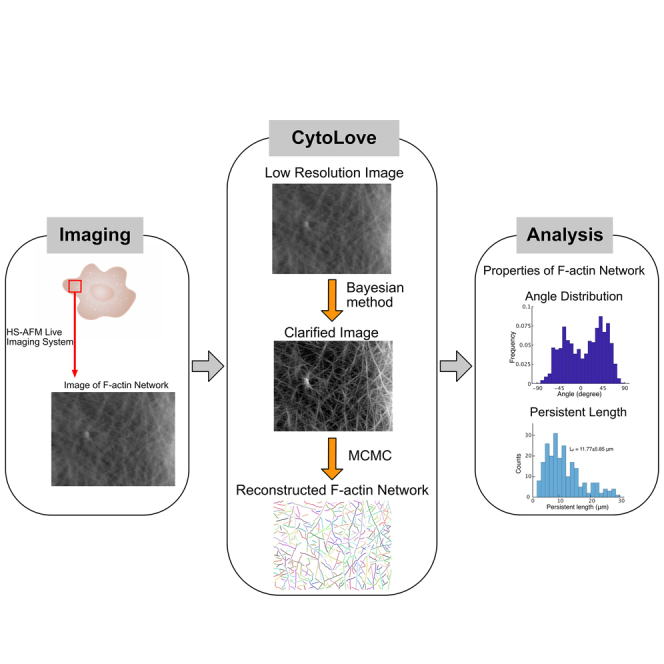
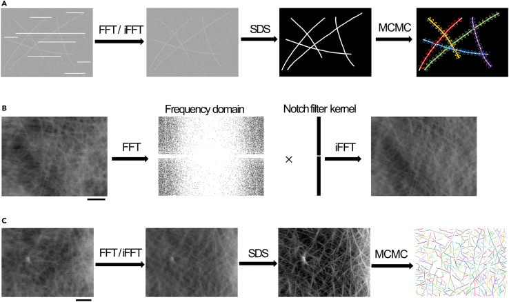
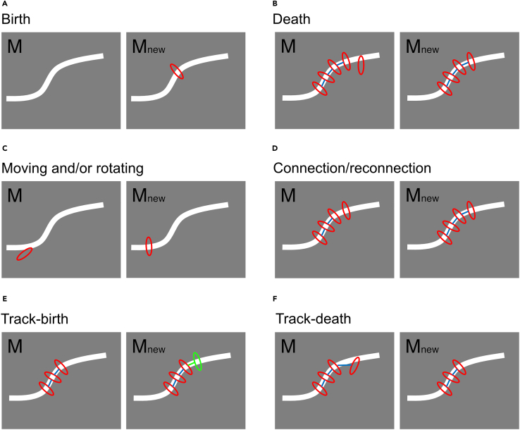
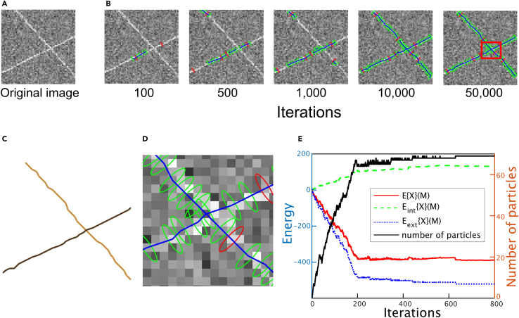
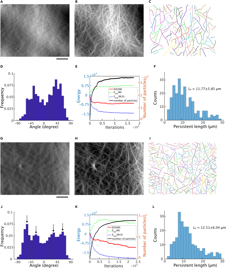
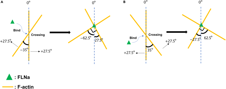
How actin filaments (F-actins) are dynamically reorganized in motile cells at the level of individual filaments is an open question. To find the answer, a high-speed atomic force microscopy (HS-AFM) system has been developed to live-imagine intracellular dynamics of the individual F-actins. However, noise and low resolution made it difficult to fully recognize individual F-actins in the HS-AFM images. To tackle this problem, we developed a new machine learning method that quantitatively recognizes individual F-actins. The method estimates F-actin orientation from the image while improving the resolution. We found that F-actins were oriented at ±35° toward the membrane in the lamellipodia, which is consistent with Arp2/3 complex-induced branching. Furthermore, in the cell cortex our results showed non-random orientation at four specific angles, suggesting a new mechanism for F-actin organization demonstrating the potential of our newly developed method to fundamentally improve our understanding of the structural dynamics of F-actin networks.
在运动细胞中,肌动蛋白丝(F-肌动蛋白)如何在单根丝的水平上进行动态重组仍是一个悬而未决的问题。为了找到答案,人们开发了一种高速原子力显微镜(HS-AFM)系统,以实时成像单个F-肌动蛋白的细胞内动态。然而,噪声和低分辨率使得在HS-AFM图像中难以完全识别单个F-肌动蛋白。为了解决这个问题,我们开发了一种新的机器学习方法,用于定量识别单个F-肌动蛋白。该方法在提高分辨率的同时,从图像中估计F-肌动蛋白的方向。我们发现,在片状伪足中,F-肌动蛋白以±35°的角度朝向细胞膜定向,这与Arp2/3复合物诱导的分支一致。此外,在细胞皮层中,我们的结果显示在四个特定角度存在非随机定向,这表明了一种F-肌动蛋白组织的新机制,证明了我们新开发的方法在从根本上提高我们对F-肌动蛋白网络结构动态理解方面的潜力。