Chugh Priyamvada, Clark Andrew G, Smith Matthew B, Cassani Davide A D, Dierkes Kai, Ragab Anan, Roux Philippe P, Charras Guillaume, Salbreux Guillaume, Paluch Ewa K
MRC Laboratory for Molecular Cell Biology, University College London, London WC1E 6BT, UK.
Centre for Genomic Regulation, The Barcelona Institute of Science and Technology, Barcelona 08003, Spain.
Nat Cell Biol. 2017 Jun;19(6):689-697. doi: 10.1038/ncb3525. Epub 2017 May 22.
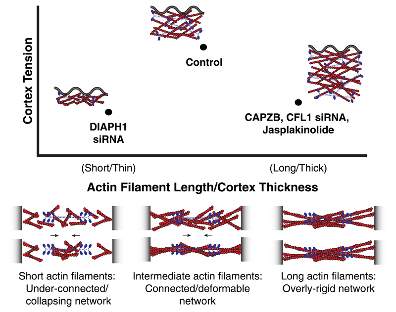
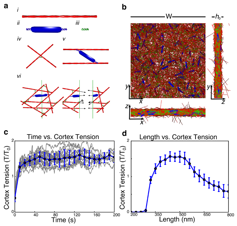
Animal cell shape is largely determined by the cortex, a thin actin network underlying the plasma membrane in which myosin-driven stresses generate contractile tension. Tension gradients result in local contractions and drive cell deformations. Previous cortical tension regulation studies have focused on myosin motors. Here, we show that cortical actin network architecture is equally important. First, we observe that actin cortex thickness and tension are inversely correlated during cell-cycle progression. We then show that the actin filament length regulators CFL1, CAPZB and DIAPH1 regulate mitotic cortex thickness and find that both increasing and decreasing thickness decreases tension in mitosis. This suggests that the mitotic cortex is poised close to a tension maximum. Finally, using a computational model, we identify a physical mechanism by which maximum tension is achieved at intermediate actin filament lengths. Our results indicate that actin network architecture, alongside myosin activity, is key to cell surface tension regulation.
动物细胞的形状很大程度上由皮层决定,皮层是质膜下的一层薄肌动蛋白网络,其中肌球蛋白驱动的应力产生收缩张力。张力梯度导致局部收缩并驱动细胞变形。先前关于皮层张力调节的研究主要集中在肌球蛋白马达上。在这里,我们表明皮层肌动蛋白网络结构同样重要。首先,我们观察到在细胞周期进程中,肌动蛋白皮层厚度与张力呈负相关。然后我们表明,肌动蛋白丝长度调节因子CFL1、CAPZB和DIAPH1调节有丝分裂皮层厚度,并发现增加和减少厚度都会降低有丝分裂中的张力。这表明有丝分裂皮层处于接近最大张力的状态。最后,我们使用一个计算模型,确定了一种在中等肌动蛋白丝长度下实现最大张力的物理机制。我们的结果表明,肌动蛋白网络结构与肌球蛋白活性一起,是细胞表面张力调节的关键。