Institute of Zoology / Developmental Biology, University of Cologne, Cologne, Germany.
Center for Molecular Medicine Cologne (CMMC), University of Cologne, Cologne, Germany.
Cell Death Dis. 2024 Oct 14;15(10):746. doi: 10.1038/s41419-024-07134-2.
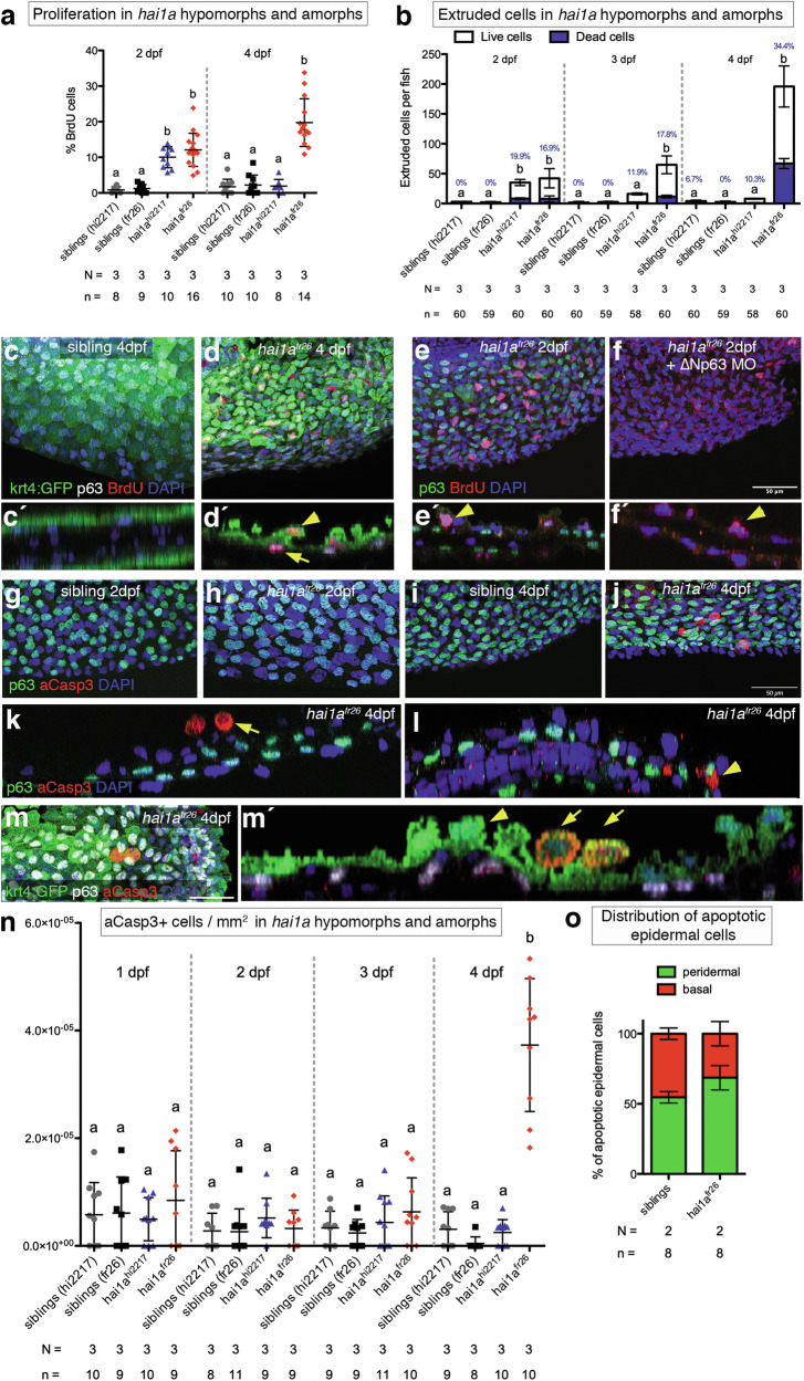
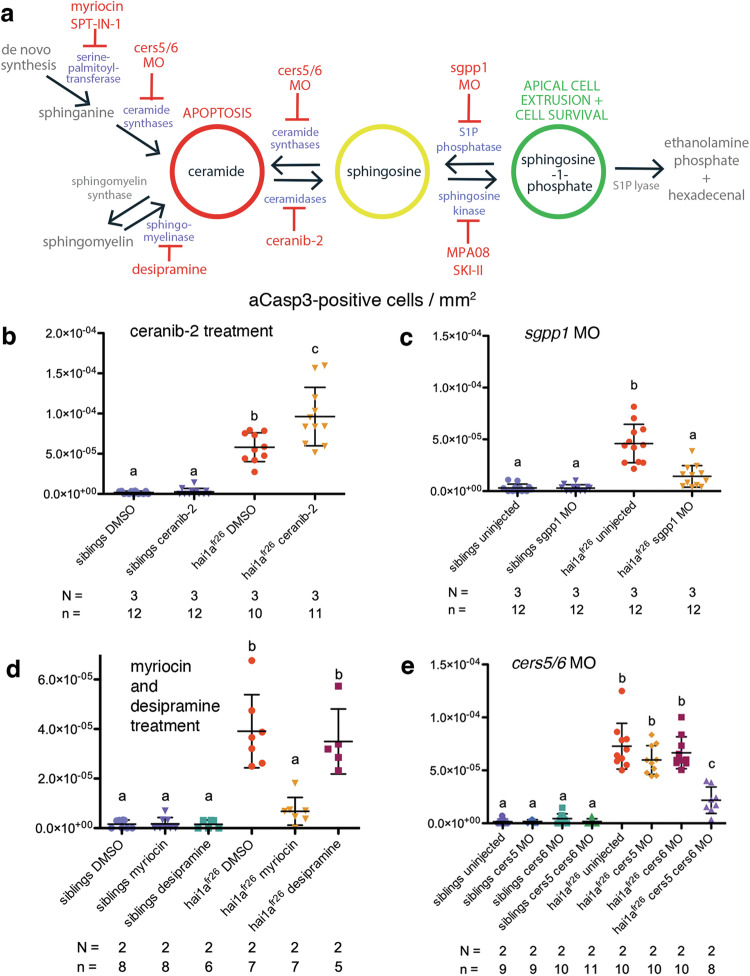
Evasion of cell death is a hallmark of cancer, and consequently the induction of cell death is a common strategy in cancer treatment. However, the molecular mechanisms regulating different types of cell death are poorly understood. We have formerly shown that in the epidermis of hypomorphic zebrafish hai1a mutant embryos, pre-neoplastic transformations of keratinocytes caused by unrestrained activity of the type II transmembrane serine protease Matriptase-1 heal spontaneously. This healing is driven by Matriptase-dependent increased sphingosine kinase (SphK) activity and sphingosine-1-phosphate (S1P)-mediated keratinocyte loss via apical cell extrusion. In contrast, amorphic hai1a mutants with even higher Matriptase-1 and SphK activity die within a few days. Here we show that this lethality is not due to epidermal carcinogenesis, but to aberrant tp53-independent apoptosis of keratinocytes caused by increased levels of pro-apoptotic C ceramides, sphingolipid counterparts to S1P within the sphingolipid rheostat, which severely compromises the epidermal barrier. Mathematical modelling of sphingolipid rheostat homeostasis, combined with in vivo manipulations of components of the rheostat or the ceramide de novo synthesis pathway, indicate that this unexpected overproduction of ceramides is caused by a negative feedback loop sensing ceramide levels and controlling ceramide replenishment via de novo synthesis. Therefore, despite their initial decrease due to increased conversion to S1P, ceramides eventually reach cell death-inducing levels, making transformed pre-neoplastic keratinocytes die even before they are extruded, thereby abrogating the normally barrier-preserving mode of apical live cell extrusion. Our results offer an in vivo perspective of the dynamics of sphingolipid homeostasis and its relevance for epithelial cell survival versus cell death, linking apical cell extrusion and apoptosis. Implications for human carcinomas and their treatments are discussed.
细胞死亡逃避是癌症的一个标志,因此诱导细胞死亡是癌症治疗的常见策略。然而,调节不同类型细胞死亡的分子机制还了解甚少。我们之前曾表明,在功能减弱的 zebrafish hai1a 突变体胚胎的表皮中,由于 II 型跨膜丝氨酸蛋白酶 Matriptase-1 的不受控制的活性导致的角质形成细胞前肿瘤转化会自发愈合。这种愈合是由 Matriptase 依赖性增加的鞘氨醇激酶 (SphK) 活性和 SphK 依赖性增加驱动的,导致 SphK 活性增加,从而通过顶端细胞外排导致鞘氨醇-1-磷酸 (S1P) 介导的角质形成细胞丢失。相比之下,具有更高 Matriptase-1 和 SphK 活性的无功能 hai1a 突变体在几天内死亡。在这里,我们表明这种致死性不是由于表皮癌变,而是由于角质形成细胞中促凋亡 C ceramides 的水平增加导致的异常 tp53 非依赖性细胞凋亡,sphingolipid rheostat 中的 sphingolipid rheostat 的 SphK 活性增加,这严重损害了表皮屏障。稳态,结合 sphingolipid rheostat 稳态的数学建模,以及对 rheostat 或 ceramide de novo 合成途径成分的体内操作,表明这种 ceramide 产量的意外增加是由一种负反馈回路引起的,该回路通过从头合成来感知 ceramide 水平并控制 ceramide 补充。因此,尽管由于转化为 S1P 而最初减少,但 ceramides 最终达到诱导细胞死亡的水平,使得转化的前肿瘤角质形成细胞甚至在被挤出之前就死亡,从而破坏了正常的顶端活细胞挤出的屏障保留模式。我们的结果提供了 sphingolipid 稳态动力学的体内视角及其对上皮细胞存活与细胞死亡的相关性,将顶端细胞外排与细胞凋亡联系起来。讨论了其对人类癌和治疗的影响。