Aramberri Jaime, Lauzirika Gorka, Illarramendi Igor, Mendicute Javier
Anterior Segment Department, Miranza Begitek, Donostia-San Sebastián, Spain.
Anterior Segment Department, Miranza Ókular, Vitoria-Gasteiz, Spain.
Clin Ophthalmol. 2024 Oct 9;18:2831-2841. doi: 10.2147/OPTH.S487627. eCollection 2024.
To compare corneal aberrometry, densitometry, and refractive outcomes of single-step Transepithelial Photorefractive Keratectomy (Trans-PRK) with and without epithelial thickness customization.
This was a prospective, interventional, randomized controlled study. Patients undergoing Trans-PRK using the WaveLight EX500 laser with StreamLight software (Alcon Laboratories, Forth Worth, TX, USA) were randomly assigned to control (55 µm standard epithelial thickness) or customized (thinnest point of epithelial thickness for each patient) groups. MS-39 (CSO, Italy) anterior segment optical coherence tomography was used to measure the epithelial thickness. Inclusion criteria were spherical equivalent <6 diopters (D), astigmatism <4D, and CDVA 20/25 or better. The assessments were at baseline and 6 months post-op: visual acuity, refraction, aberrometry, and corneal densitometry.
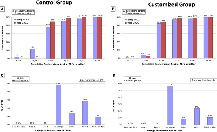
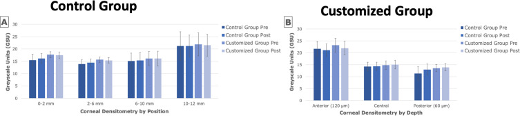
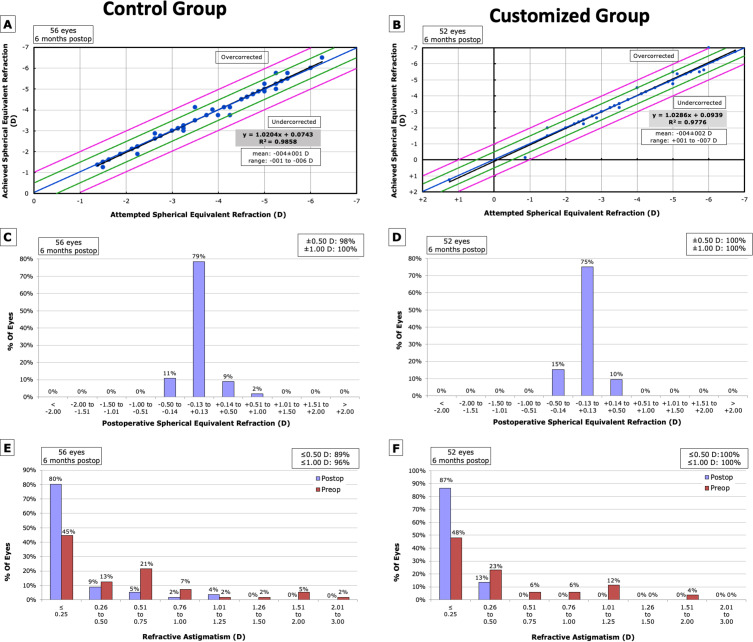
108 eyes were enrolled, [control group (n=56) and customized group (n=52)]. Mean epithelial ablation thickness in the customized group was 54.81±3.56µm (p=0.470 vs control group). Both groups experienced significant postoperative increases in higher-order aberrations (HOA) and spherical aberrations, with no significant intergroup differences. Mean HOA RMS (µm) of the frontal cornea and total cornea increased by 0.27, and 0.29, respectively, in the control group, and 0.26 and 0.28, respectively, in the customized group (p<0.001 for all). Mean change in spherical aberrations in the frontal cornea and total cornea was 0.23µm (p<0.001) and 0.25µm (p<0.001), in the control group, and 0.19µm (p<0.001) and 0.20µm (p<0.001), in the customized group. Mean corneal densitometry in anterior cornea decreased by 0.63GSU (p=0.021) and 1.18GSU (p<0.001) in the control and customized groups. In the posterior cornea, it increased by 1.67GSU (p=0.004) and 0.38GSU (p=0.006).
No significant differences in refractive and aberrometry outcomes between control and customized Trans-PRK groups, with corneal densitometry changes not affecting visual acuity.
比较单步经上皮光屈光性角膜切削术(Trans-PRK)在有无上皮厚度个体化定制情况下的角膜像差测量、密度测量及屈光结果。
这是一项前瞻性、干预性、随机对照研究。使用配备StreamLight软件的威视EX500激光(爱尔康实验室,美国得克萨斯州沃思堡)进行Trans-PRK的患者被随机分为对照组(标准上皮厚度55µm)或个体化定制组(每位患者上皮厚度最薄处)。采用MS-39(意大利CSO)眼前节光学相干断层扫描测量上皮厚度。纳入标准为等效球镜度<6屈光度(D)、散光<4D且矫正远视力(CDVA)为20/25或更佳。评估在基线及术后6个月进行:视力、验光、像差测量及角膜密度测量。
共纳入108只眼,[对照组(n = 56)和个体化定制组(n = 52)]。个体化定制组的平均上皮切削厚度为54.81±3.56µm(与对照组相比,p = 0.470)。两组术后高阶像差(HOA)和球差均显著增加,组间无显著差异。对照组角膜前表面和全角膜的平均HOA均方根(µm)分别增加0.27和0.29,个体化定制组分别增加0.26和0.28(所有p<0.001)。对照组角膜前表面和全角膜的球差平均变化分别为0.23µm(p<0.001)和0.25µm(p<0.001),个体化定制组分别为0.19µm(p<0.001)和0.20µm(p<0.001)。对照组和个体化定制组角膜前表面的平均角膜密度测量值分别降低0.63GSU(p = 0.021)和1.18GSU(p<0.001)。在角膜后表面,其分别增加1.67GSU(p = 0.004)和0.38GSU(p = 0.006)。
对照组和个体化定制Trans-PRK组在屈光和像差测量结果上无显著差异,角膜密度测量变化不影响视力。