Department of Applied Biochemistry, Institute of Biotechnology, Technische Universität Berlin, 10623 Berlin, Germany.
Department of Radiation Oncology, Charité University Medicine Berlin, Corporate Member of Freie Universität Berlin and Humboldt-Universität Zu Berlin, 13353 Berlin, Germany.
Int J Mol Sci. 2024 Sep 24;25(19):10268. doi: 10.3390/ijms251910268.
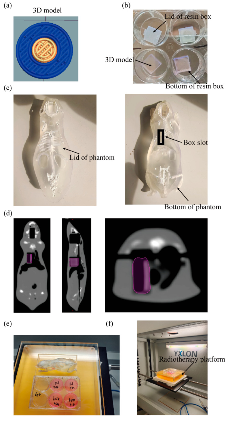
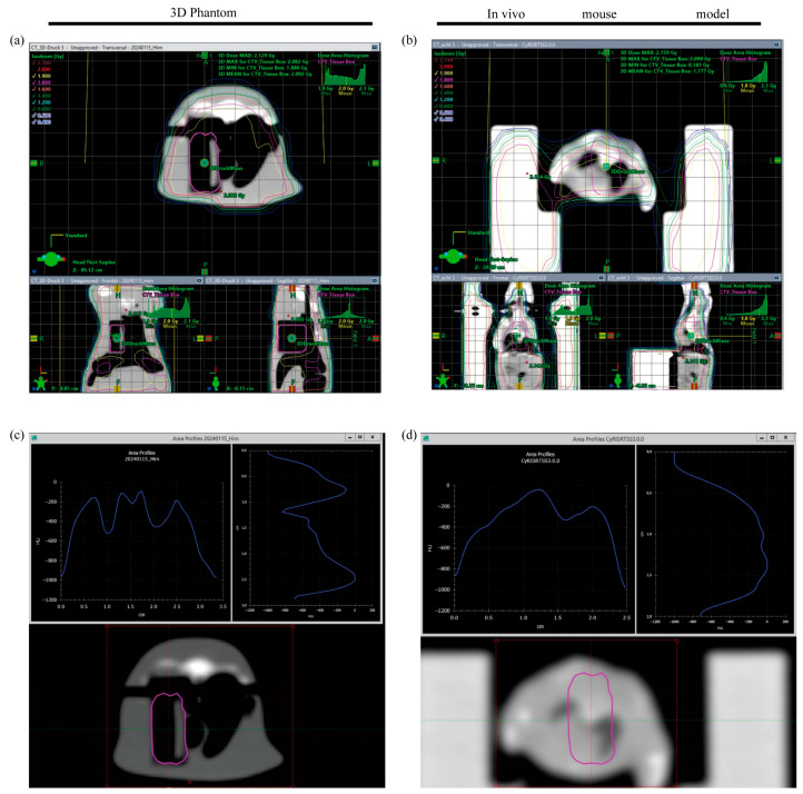
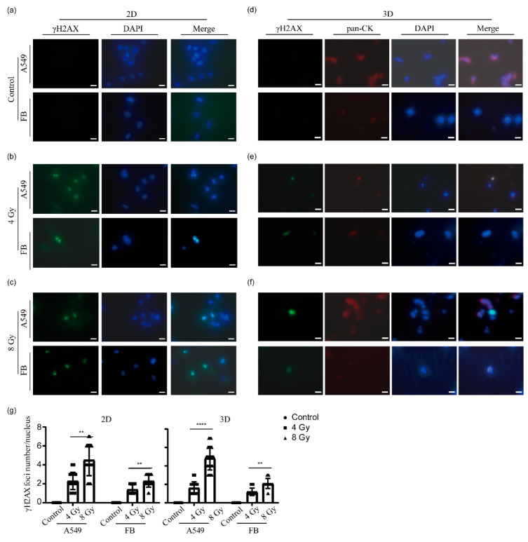
Lung cancer continues to have one of the highest morbidity and mortality rates of any cancer. Although radiochemotherapy, in combination with immunotherapy, has significantly improved overall survival, new treatment options are urgently needed. However, preclinical radiotherapy testing is often performed in animal models, which has several drawbacks, including species-specific differences and ethical concerns. To replace animal models, this study used a micro-extrusion bioprinting approach to generate a three-dimensional (3D) human lung cancer model consisting of lung tumor cells embedded in human primary lung fibroblasts for radiotherapy research. The models were placed in a mouse phantom, i.e., a 3D-printed mouse model made of materials that mimic the X-ray radiation attenuation rates found in mice. In radiotherapy experiments, the model demonstrated a selective cytotoxic effect of X-rays on tumor cells, consistent with findings in 2D cells. Furthermore, the analysis of metabolic activity, cell death, apoptosis, and DNA damage-induced γH2AX foci formation revealed different results in the 3D model inside the phantom compared to those observed in irradiated models without phantom and 2D cells. The proposed setup of the bioprinted 3D lung model inside the mouse phantom provides a physiologically relevant model system to study radiation effects.
肺癌仍然是发病率和死亡率最高的癌症之一。虽然放化疗联合免疫疗法显著提高了总生存率,但仍迫切需要新的治疗选择。然而,临床前放射治疗试验通常在动物模型中进行,这存在几个缺点,包括物种特异性差异和伦理问题。为了替代动物模型,本研究采用微挤出生物打印方法生成了一个由嵌入人原代肺成纤维细胞中的肺癌细胞组成的三维(3D)人类肺癌模型,用于放射治疗研究。将模型放置在小鼠体模中,即由模拟小鼠中 X 射线辐射衰减率的材料制成的 3D 打印小鼠模型。在放射治疗实验中,该模型显示出 X 射线对肿瘤细胞的选择性细胞毒性作用,与 2D 细胞的结果一致。此外,对代谢活性、细胞死亡、细胞凋亡和 DNA 损伤诱导的 γH2AX 焦点形成的分析表明,在体模内的 3D 模型中观察到的结果与没有体模和 2D 细胞的照射模型以及 2D 细胞观察到的结果不同。该生物打印的 3D 肺模型在小鼠体模内部的构建为研究辐射效应提供了一个具有生理相关性的模型系统。