Department of Applied Biochemistry, Institute of Biotechnology, Technische Universität Berlin, 13355 Berlin, Germany.
Department of Radiooncology, Rostock University Medical Center, 18059 Rostock, Germany.
Int J Mol Sci. 2022 Sep 1;23(17):9951. doi: 10.3390/ijms23179951.
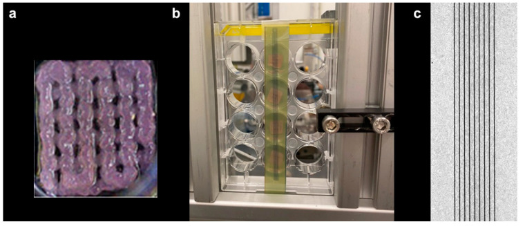
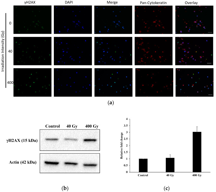
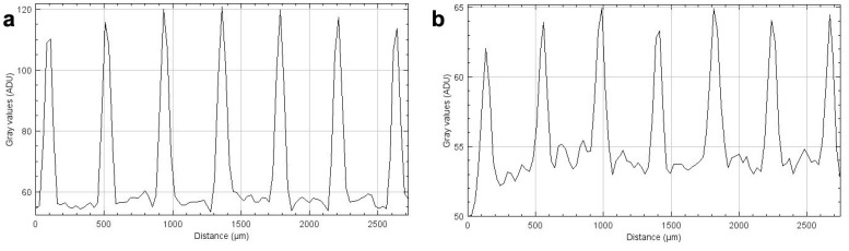
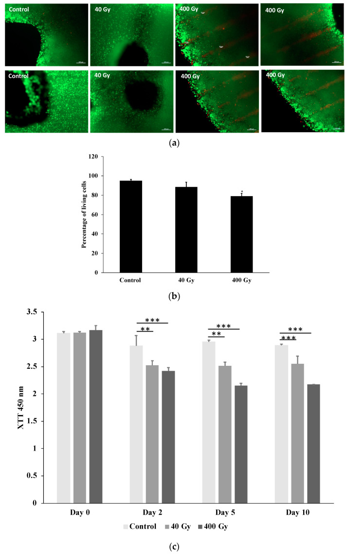
Radiotherapy is an important component in the treatment of lung cancer, one of the most common cancers worldwide, frequently resulting in death within only a few years of diagnosis. In order to evaluate new therapeutic approaches and compare their efficiency with regard to tumour control at a pre-clinical stage, it is important to develop standardized samples which can serve as inter-institutional outcome controls, independent of differences in local technical parameters or specific techniques. Recent developments in 3D bioprinting techniques could provide a sophisticated solution to this challenge. We have conducted a pilot project to evaluate the suitability of standardized samples generated from 3D printed human lung cancer cells in radiotherapy studies. The samples were irradiated at high dose rates using both broad beam and microbeam techniques. We found the 3D printed constructs to be sufficiently mechanically stable for use in microbeam studies with peak doses up to 400 Gy to test for cytotoxicity, DNA damage, and cancer cell death in vitro. The results of this study show how 3D structures generated from human lung cancer cells in an additive printing process can be used to study the effects of radiotherapy in a standardized manner.
放射治疗是治疗肺癌的重要组成部分,肺癌是全球最常见的癌症之一,通常在诊断后仅几年内就会导致死亡。为了在临床前阶段评估新的治疗方法并比较它们在肿瘤控制方面的效率,开发可作为机构间结果对照的标准化样本非常重要,这些样本不受局部技术参数或特定技术差异的影响。最近 3D 生物打印技术的发展为此提供了一个复杂的解决方案。我们进行了一个试点项目,以评估 3D 打印的人类肺癌细胞生成的标准化样本在放射治疗研究中的适用性。使用宽束和微束技术以高剂量率对样本进行照射。我们发现 3D 打印结构足够稳定,可以用于微束研究,峰值剂量高达 400Gy,以测试体外细胞毒性、DNA 损伤和癌细胞死亡。这项研究的结果表明,如何使用增材打印工艺生成的源自人类肺癌细胞的 3D 结构可用于以标准化方式研究放射治疗的效果。