Bakker Jens T, Dudurych Ivan, Roodenburg Sharyn A, Vonk Judith M, Klooster Karin, de Bruijne Marleen, van den Berge Maarten, Slebos Dirk-Jan, Vliegenthart Rozemarijn
Department of Pulmonary Diseases, University of Groningen, University Medical Center Groningen, Groningen, The Netherlands.
Groningen Research Institute for Asthma and COPD (GRIAC), University of Groningen, University Medical Center Groningen, Groningen, The Netherlands.
Eur Radiol. 2025 May;35(5):2912-2921. doi: 10.1007/s00330-024-11123-6. Epub 2024 Oct 16.
Lung hyperinflation, a key contributor to dyspnea in chronic obstructive pulmonary disease (COPD), can be quantified via chest computed tomography (CT). Establishing reference equations for lobar volumes and total lung volume (TLV) can aid in evaluating lobar hyperinflation, especially for targeted lung volume reduction therapies.
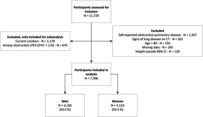
The Imaging in Lifelines study (ImaLife) comprises 11,729 participants aged 45 and above with analyzed inspiratory low-dose thoracic CT scans. Lung and lobar volumes were measured using an automatic AI-based segmentation algorithm (LungSeg). For the main analysis, participants were excluded if they had self-reported COPD/asthma, lung disease on CT, airflow obstruction on lung function testing, were currently smoking, aged over 80 years, or had height outside the 99% confidence interval. Reference equations for TLV and lobar volumes were determined using linear regression considering age and height, stratified by sex. For the subanalysis, participants who were currently smoking or experiencing airflow obstruction were compared to the group of the main analysis.
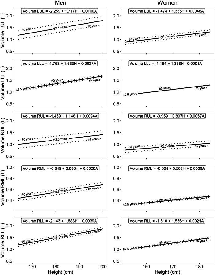
The study included 7306 lung-healthy participants, 97.5% Caucasian, 43.6% men, with mean age of 60.3 ± 9.5 years. Lung and lobar volumes generally increased with age and height. Men consistently had higher volumes than women when adjusted for height. R values ranged from 7.8 to 19.9%. In smokers and those with airway obstruction, volumes were larger than in lung-healthy groups, with the largest increases measured in the upper lobes.
The established reference equations for CT-derived TLV and lobar volumes provide a standardized interpretation for individuals aged 45 to 80 of Northern European descent.
Question Lobar lung volumes can be derived from inspiratory CT scans, but healthy-lung reference values are lacking. Findings Lung and lobar volumes generally increased with age and height. Reference equations for lung/lobar volumes were derived from a sizeable lung-healthy population. Clinical relevance This study provides reference equations for inspiratory CT-derived lung and lobar volumes in a lung-healthy population, potentially useful for assessing candidates for lung volume reduction therapies, for lobe removal in lung cancer patients, and in case of restrictive pulmonary diseases.
肺过度充气是慢性阻塞性肺疾病(COPD)患者呼吸困难的关键因素,可通过胸部计算机断层扫描(CT)进行量化。建立叶体积和总肺容量(TLV)的参考方程有助于评估叶过度充气,特别是对于有针对性的肺减容治疗。
生命线成像研究(ImaLife)纳入了11729名年龄在45岁及以上的参与者,并对其吸气期低剂量胸部CT扫描进行了分析。使用基于人工智能的自动分割算法(LungSeg)测量肺和叶体积。在主要分析中,如果参与者自我报告有COPD/哮喘、CT显示肺部疾病、肺功能测试存在气流阻塞、当前正在吸烟、年龄超过80岁或身高超出99%置信区间,则将其排除。考虑年龄和身高,通过线性回归确定TLV和叶体积的参考方程,并按性别分层。在亚分析中,将当前吸烟或存在气流阻塞的参与者与主要分析组进行比较。
该研究纳入了7306名肺部健康的参与者,其中97.5%为白种人,43.6%为男性,平均年龄为60.3±9.5岁。肺和叶体积一般随年龄和身高增加。调整身高后,男性的体积始终高于女性。R值范围为7.8%至19.9%。吸烟者和气道阻塞者的体积大于肺部健康组,上叶增加最为明显。
所建立的基于CT的TLV和叶体积参考方程为北欧血统、年龄在45至80岁的个体提供了标准化的解释。
问题 叶肺体积可从吸气期CT扫描得出,但缺乏健康肺的参考值。发现 肺和叶体积一般随年龄和身高增加。肺/叶体积的参考方程来自大量肺部健康人群。临床意义 本研究为肺部健康人群吸气期CT得出的肺和叶体积提供了参考方程,可能有助于评估肺减容治疗的候选者、肺癌患者的肺叶切除情况以及限制性肺病的情况。