Kowa Chao-Ying, Morecroft Megan, Macfarlane Alan J R, Burckett-St Laurent David, Pawa Amit, West Simeon, Margetts Steve, Haslam Nat, Ashken Toby, Sebastian Maria Paz, Thottungal Athmaja, Womack Jono, Noble Julia Alison, Higham Helen, Bowness James S
Department of Anaesthesia, The Royal London Hospital, London, UK.
Faculty of Medicine, Health & Life Sciences, University of Swansea, Swansea, UK.
BMJ Surg Interv Health Technol. 2024 Oct 16;6(1):e000264. doi: 10.1136/bmjsit-2024-000264. eCollection 2024.
Ultrasound-guided regional anesthesia (UGRA) relies on acquiring and interpreting an appropriate view of sonoanatomy. Artificial intelligence (AI) has the potential to aid this by applying a color overlay to key sonoanatomical structures.The primary aim was to determine whether an AI-generated color overlay was associated with a difference in participants' ability to identify an appropriate block view over a 2-month period after a standardized teaching session (as judged by a blinded assessor). Secondary outcomes included the ability to identify an appropriate block view (unblinded assessor), global rating score and participant confidence scores.
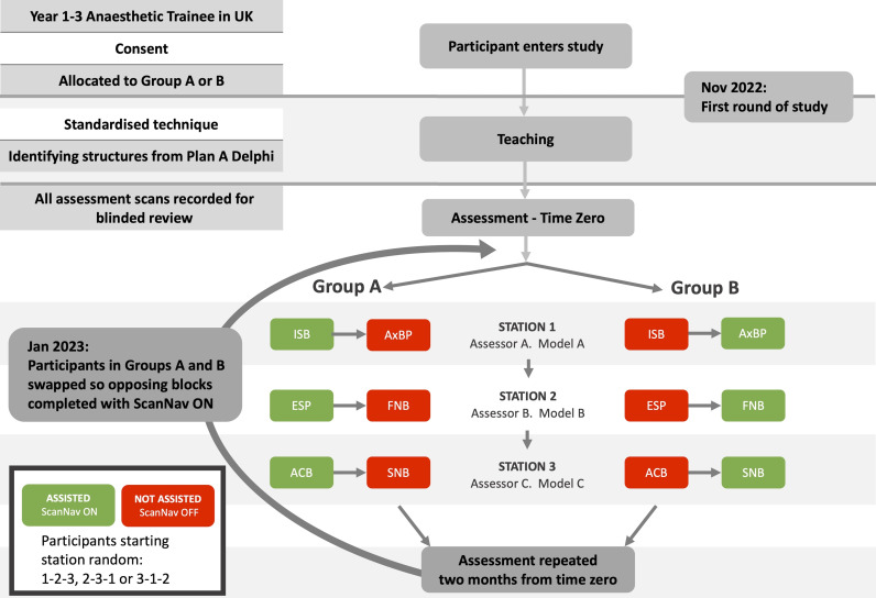
Randomized, partially blinded, prospective cross-over study.
Simulation scans on healthy volunteers. Initial assessments on 29 November 2022 and 30 November 2022, with follow-up on 25 January 2023 - 27 January 2023.
57 junior anesthetists undertook initial assessments and 51 (89.47%) returned at 2 months.
Participants performed ultrasound scans for six peripheral nerve blocks, with AI assistance randomized to half of the blocks. Cross-over assignment was employed for 2 months.
Blinded experts assessed whether the block view acquired was acceptable (yes/no). Unblinded experts also assessed this parameter and provided a global performance rating (0-100). Participants reported scan confidence (0-100).
AI assistance was associated with a higher rate of appropriate block view acquisition in both blinded and unblinded assessments (p=0.02 and <0.01, respectively). Participant confidence and expert rating scores were superior throughout (all p<0.01).
Assistive AI was associated with superior ultrasound scanning performance 2 months after formal teaching. It may aid application of sonoanatomical knowledge and skills gained in teaching, to support delivery of UGRA beyond the immediate post-teaching period.
超声引导区域麻醉(UGRA)依赖于获取并解读合适的超声解剖视图。人工智能(AI)有潜力通过为关键超声解剖结构应用颜色叠加来辅助这一过程。主要目的是确定在标准化教学课程后的2个月期间,人工智能生成的颜色叠加是否与参与者识别合适阻滞视图的能力差异相关(由盲法评估者判断)。次要结果包括识别合适阻滞视图的能力(非盲法评估者)、整体评分以及参与者信心评分。
随机、部分盲法、前瞻性交叉研究。
对健康志愿者进行模拟扫描。于2022年11月29日和2022年11月30日进行初始评估,并于2023年1月25日至2023年1月27日进行随访。
57名初级麻醉医生进行了初始评估,其中51名(89.47%)在2个月后返回。
参与者进行6次外周神经阻滞的超声扫描,人工智能辅助随机分配至一半的阻滞操作中。采用交叉分配方式持续2个月。
盲法专家评估获取的阻滞视图是否可接受(是/否)。非盲法专家也评估此参数并给出整体表现评分(0 - 100)。参与者报告扫描信心(0 - 100)。
在盲法和非盲法评估中,人工智能辅助均与更高比例的合适阻滞视图获取相关(分别为p = 0.02和p < 0.01)。参与者信心和专家评分在整个过程中均更高(所有p < 0.01)。
在正式教学2个月后,辅助性人工智能与更优的超声扫描表现相关。它可能有助于将教学中获得的超声解剖知识和技能应用于实际,以支持在教学后即时阶段之外的超声引导区域麻醉实施。