Department of Neurology, Research Institute of Neuromuscular and Neurodegenerative Diseases, Shandong Provincial Key Laboratory of Mitochondrial Medicine and Rare Diseases, Qilu Hospital of Shandong University, West Wenhua Street No.107, Jinan, 250012, Shandong, China.
Department of Radiology, Cheeloo College of Medicine, Qilu Hospital, Shandong University, Jinan, China.
Fluids Barriers CNS. 2024 Oct 21;21(1):83. doi: 10.1186/s12987-024-00586-w.
Using neuroimaging techniques, growing evidence has suggested that the choroid plexus (CP) volume is enlarged in multiple neurodegenerative diseases, including amyotrophic lateral sclerosis (ALS). Notably, the CP has been suggested to play an important role in inflammation-induced CNS damage under disease conditions. However, to our knowledge, no study has investigated the relationships between peripheral inflammation and CP volume in sporadic ALS patients. Thus, in this study, we aimed to verify CP enlargement and explore its association with peripheral inflammation in vivo in independent ALS cohorts.
Based on structural MRI data, CP volume was measured using Gaussian mixture models and further manually corrected in two independent cohorts of sporadic ALS patients and healthy controls (HCs). Serum inflammatory protein levels were measured using a novel high-sensitivity Olink proximity extension assay (PEA) technique. Xtreme gradient boosting (XGBoost) was used to explore the contribution of peripheral inflammatory factors to CP enlargement. Then, partial correlation analyses were performed.
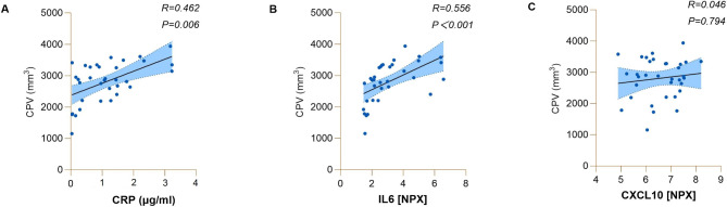
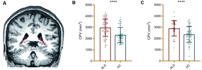
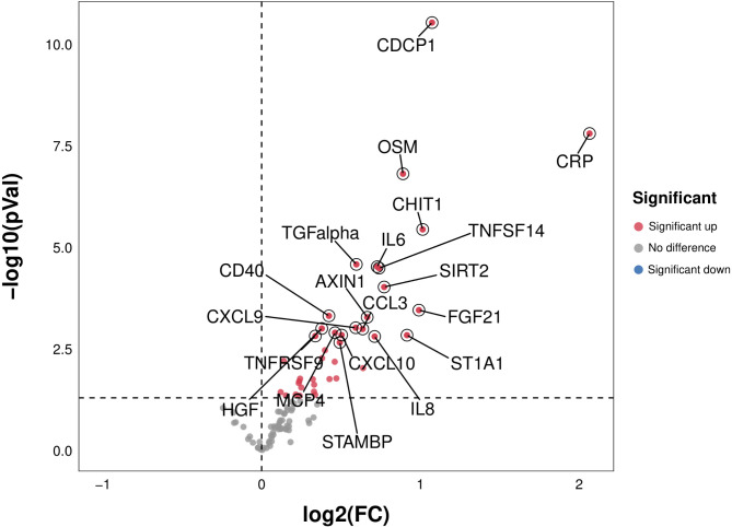
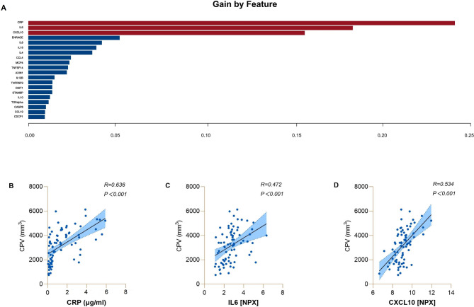
CP volumes were significantly higher in ALS patients than in HCs in the independent cohorts. Compared with HCs, serum levels of CRP, IL-6, CXCL10, and 35 other inflammatory factors were significantly increased in ALS patients. Using the XGBoost approach, we established a model-based importance of features, and the top three predictors of CP volume in ALS patients were CRP, IL-6, and CXCL10 (with gains of 0.24, 0.18, and 0.15, respectively). Correlation analyses revealed that CRP, IL-6, and CXCL10 were significantly associated with CP volume in ALS patients (r = 0.462 ∼ 0.636, p < 0.001).
Our study is the first to reveal a consistent and replicable contribution of peripheral inflammation to CP enlargement in vivo in sporadic ALS patients. Given that CP enlargement has been recently detected in other brain diseases, these findings should consider extending to other disease conditions with a peripheral inflammatory component.
利用神经影像学技术,越来越多的证据表明脉络丛(CP)体积在多种神经退行性疾病中增大,包括肌萎缩侧索硬化症(ALS)。值得注意的是,CP 在疾病状态下被认为在炎症诱导的中枢神经系统损伤中发挥重要作用。然而,据我们所知,尚无研究调查散发性 ALS 患者外周炎症与 CP 体积之间的关系。因此,在这项研究中,我们旨在验证 CP 增大,并在两个独立的散发性 ALS 患者和健康对照(HC)队列中探索其与外周炎症的关系。
基于结构 MRI 数据,使用高斯混合模型测量 CP 体积,并在两个独立的散发性 ALS 患者和健康对照(HC)队列中进一步手动校正。使用新型高灵敏度 Olink 邻近延伸分析(PEA)技术测量血清炎症蛋白水平。使用极端梯度提升(XGBoost)方法探索外周炎症因子对 CP 增大的贡献。然后进行部分相关分析。
在独立队列中,ALS 患者的 CP 体积明显高于 HC。与 HC 相比,ALS 患者的 CRP、IL-6、CXCL10 和其他 35 种炎症因子的血清水平显著升高。使用 XGBoost 方法,我们建立了基于模型的特征重要性模型,ALS 患者 CP 体积的前三个预测因子是 CRP、IL-6 和 CXCL10(增益分别为 0.24、0.18 和 0.15)。相关分析显示,CRP、IL-6 和 CXCL10 与 ALS 患者的 CP 体积显著相关(r=0.462~0.636,p<0.001)。
本研究首次揭示了外周炎症对散发性 ALS 患者 CP 增大的一致且可重复的贡献。鉴于 CP 增大最近在其他脑部疾病中被检测到,这些发现应考虑扩展到具有外周炎症成分的其他疾病状态。