Laboratory of Parasitic Diseases, National Institute of Allergy and Infectious Diseases, National Institutes of Health (NIH), Bethesda, MD, United States.
Integrated Data Science Section (IDSS), National Institute of Allergy and Infectious Diseases (NIAID), National Institutes of Health (NIH), Bethesda, MD, United States.
Front Immunol. 2024 Oct 7;15:1436818. doi: 10.3389/fimmu.2024.1436818. eCollection 2024.
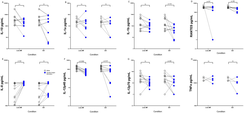
Live microfilariae (mf) and mf-derived extracellular vesicles (EVs) have been shown to modulate human antigen presenting cell (APC) function, most notably by suppressing the induction of IL-12 (and other pro-inflammatory cytokines) following activation with LPS and interferon-y.
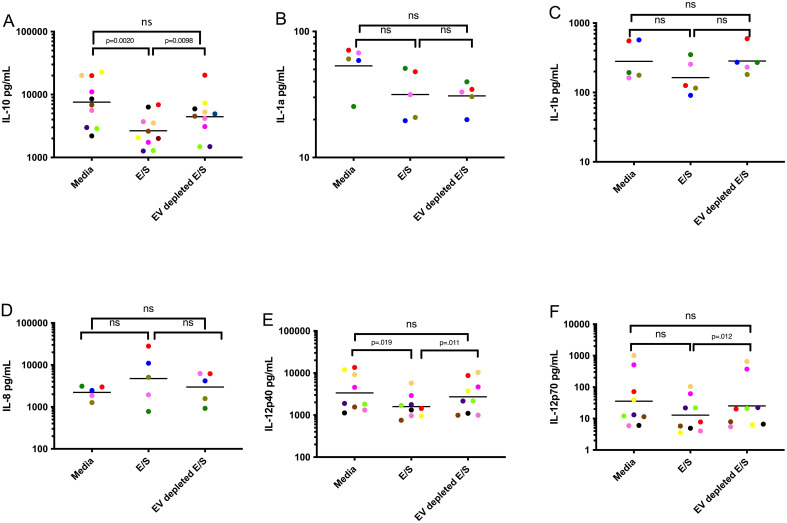
To explore further how EVs alter human APC function, we studied the effect of mf and EVs on human elutriated monocyte-derived dendritic cells (DC) following exposure to Mf, mf-derived excretory/secretory (E/S) products, E/S depleted of EVs through ultracentrifugation and purified EVs. After demonstrating that the measurable responses induced by live mf could be recapitulated by EVs and EV-containing E/S, we next performed RNAseq analysis of human DC following exposure to live mf, EVs, E/S, or EV-depleted E/S.
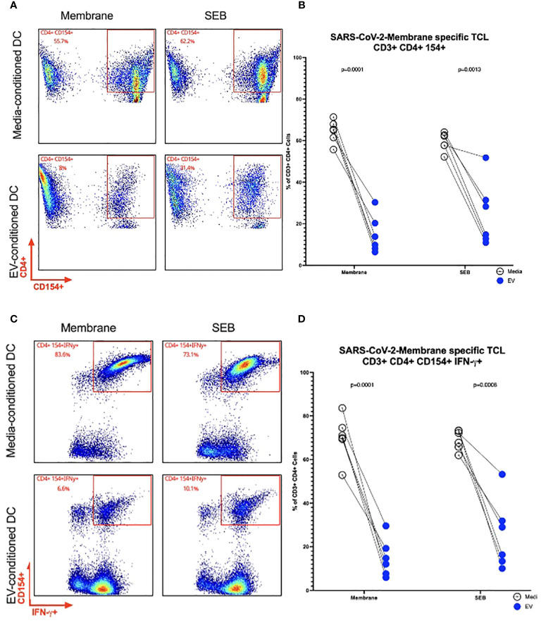
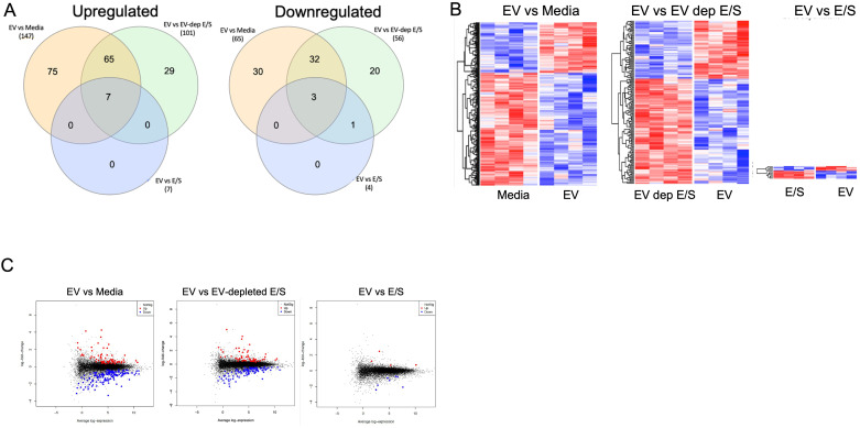
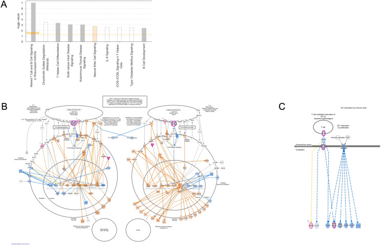
In our analyses of the data for the DC, using a false discovery rate (FDR)<0.05, EV-exposed DC had induced the expression of 212 differentially expressed genes (DEGs) when compared to unexposed DC and 157 when compared to E/S-depleted EVs. These genes were enriched in GO biological processes associated with neutrophil degranulation and 15 DEGs associated with KEGG Lysosome pathways. IPA analysis point to immune dysregulation. We next aimed to understand the intracellular processes altered by EVs and the effect these have on effector T cells. When SARS CoV-2 Membrane-specific CD4+ TCLs were assessed following EV conditioning of autologous DC and activation with the SARS CoV-2-Membrane peptide pool, we found conditioning reduced the frequency of SARS CoV-2 Membrane-specific CD3+ CD4+ CD154+ cells (p=.015). Similarly, EV-conditioning of SARS CoV-2 Membrane-specific CD3+ CD4+ cells induced fewer cell capable of producing IFN-γ (p=.045).
Taken together, our data suggest a modulatory role of EVs on APC function that likely leads to defects in T cell effector function.
已经表明,活微丝蚴(mf)和 mf 衍生的细胞外囊泡(EVs)可调节人类抗原呈递细胞(APC)的功能,特别是通过抑制 LPS 和干扰素-y 激活后 IL-12(和其他促炎细胞因子)的诱导。
为了进一步探索 EVs 如何改变人类 APC 的功能,我们研究了 mf 和 EVs 对人白细胞分离单核细胞衍生树突状细胞(DC)的影响,这些 DC 暴露于 mf、mf 衍生的外泌体/分泌产物(E/S)、通过超速离心去除 EVs 的 E/S 以及纯化的 EVs。在证明活 mf 诱导的可测量反应可通过 EV 和含 EV 的 E/S 重现后,我们接下来对暴露于活 mf、EV、E/S 或 EV 耗尽的 E/S 后的人 DC 进行了 RNAseq 分析。
在我们对 DC 数据的分析中,使用错误发现率(FDR)<0.05,与未暴露的 DC 相比,EV 暴露的 DC 诱导了 212 个差异表达基因(DEGs)的表达,与 E/S 耗尽的 EV 相比,诱导了 157 个 DEGs 的表达。这些基因在与中性粒细胞脱颗粒相关的 GO 生物学过程和与 KEGG 溶酶体途径相关的 15 个 DEGs 中富集。IPA 分析表明免疫失调。接下来,我们旨在了解 EV 改变的细胞内过程以及这些过程对效应 T 细胞的影响。当 SARS CoV-2 膜特异性 CD4+ TCL 在自体 DC 条件下并使用 SARS CoV-2 膜肽池激活后评估 EV 时,我们发现条件降低了 SARS CoV-2 膜特异性 CD3+ CD4+ CD154+细胞的频率(p=.015)。同样,EV 对 SARS CoV-2 膜特异性 CD3+ CD4+细胞的处理诱导产生 IFN-γ 的细胞减少(p=.045)。
综上所述,我们的数据表明 EVs 对 APC 功能具有调节作用,这可能导致 T 细胞效应功能缺陷。