• 文献检索
  • 文档翻译
  • 深度研究
  • 学术资讯
  • Suppr Zotero 插件Zotero 插件
  • 邀请有礼
  • 套餐&价格
  • 历史记录
应用&插件
Suppr Zotero 插件Zotero 插件浏览器插件Mac 客户端Windows 客户端微信小程序
定价
高级版会员购买积分包购买API积分包
服务
文献检索文档翻译深度研究API 文档MCP 服务
关于我们
关于 Suppr公司介绍联系我们用户协议隐私条款
关注我们

Suppr 超能文献

核心技术专利:CN118964589B侵权必究
粤ICP备2023148730 号-1Suppr @ 2026

文献检索

告别复杂PubMed语法,用中文像聊天一样搜索,搜遍4000万医学文献。AI智能推荐,让科研检索更轻松。

立即免费搜索

文件翻译

保留排版,准确专业,支持PDF/Word/PPT等文件格式,支持 12+语言互译。

免费翻译文档

深度研究

AI帮你快速写综述,25分钟生成高质量综述,智能提取关键信息,辅助科研写作。

立即免费体验

液滴微涡旋实现活悬浮细胞的三维各向同性成像。

Three-dimensional isotropic imaging of live suspension cells enabled by droplet microvortices.

机构信息

Department of Biomedical Engineering, University of California, Irvine, CA 92697.

Center for Advanced Design & Manufacturing of Integrated Microfluidics, University of California, Irvine, CA 92697.

出版信息

Proc Natl Acad Sci U S A. 2024 Oct 29;121(44):e2408567121. doi: 10.1073/pnas.2408567121. Epub 2024 Oct 22.

DOI:10.1073/pnas.2408567121
PMID:39436653
原文链接:https://pmc.ncbi.nlm.nih.gov/articles/PMC11536124/
Abstract

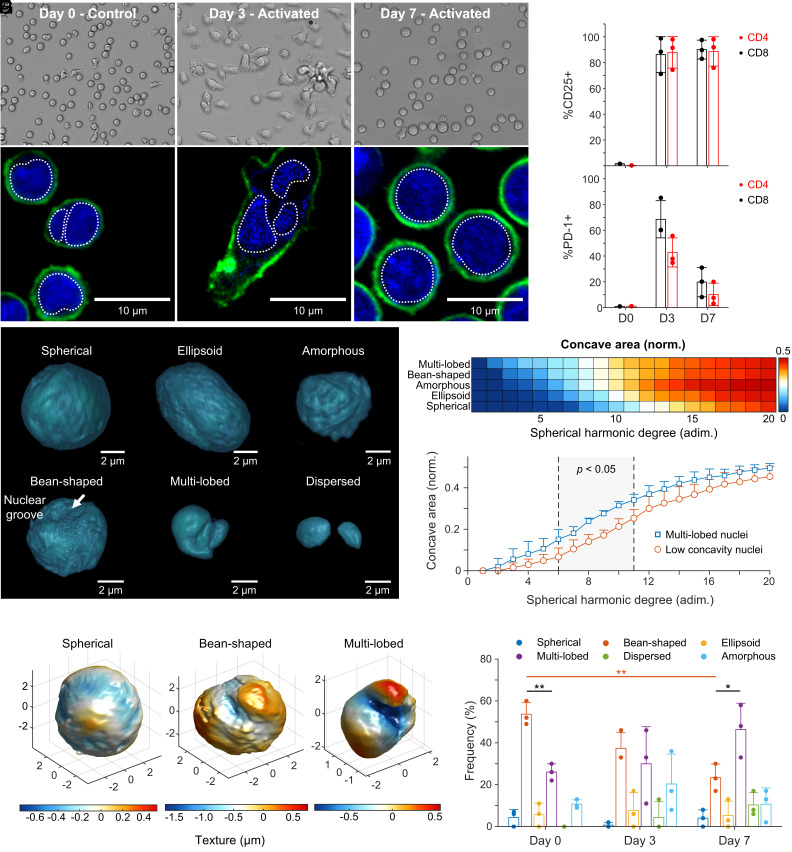
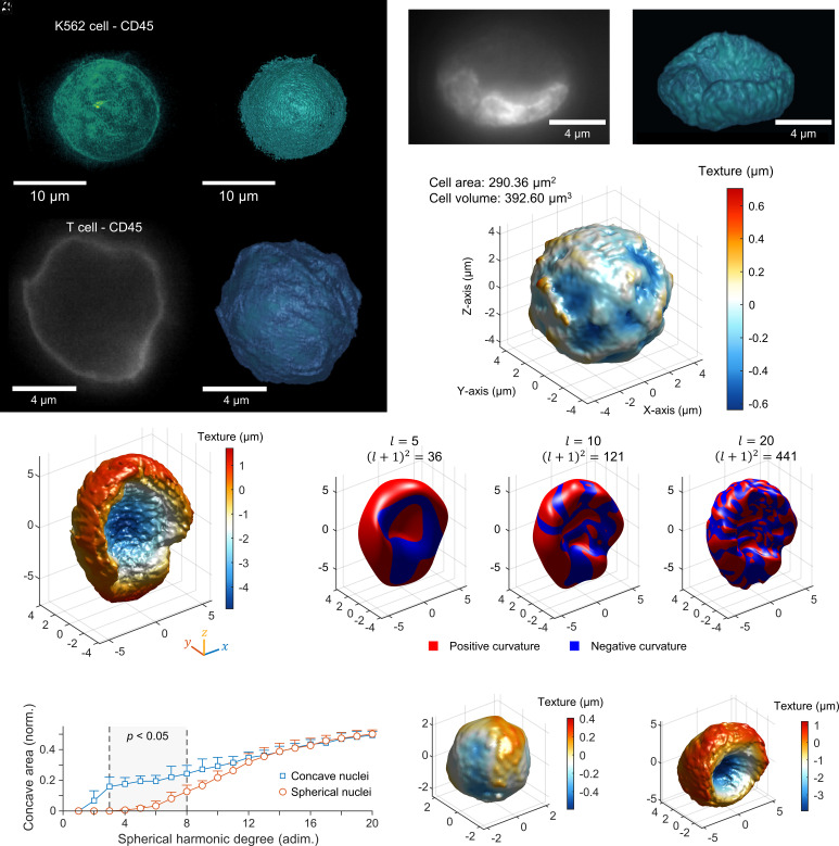
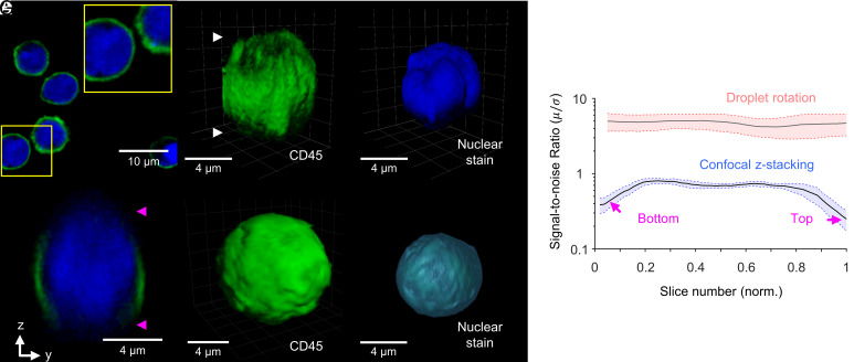
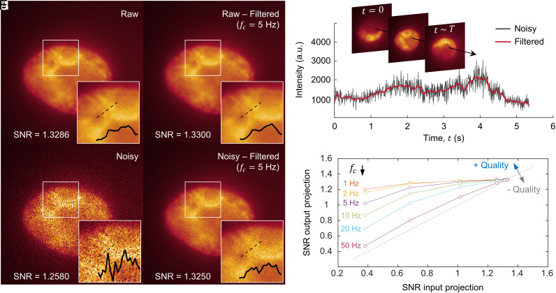
Fast, nondestructive three-dimensional (3D) imaging of live suspension cells remains challenging without substrate treatment or fixation, precluding scalable single-cell morphometry with minimal alterations. While optical sectioning techniques achieve 3D live cell imaging, lateral versus depth resolution differences further complicate analysis. We present a scalable microfluidic method capable of 3D fluorescent isotropic imaging of live, nonadherent cells suspended inside picoliter droplets with high-speed single-cell volumetric readout (800 to 1,200 slices in 5 to 8 s) and near-diffraction limit resolution (216 nm). The platform features a droplet trap array that leverages flow-induced droplet interfacial shear to generate intradroplet microvortices, which rotate single cells on their axis to enable optical projection tomography (OPT)-based imaging. This allows gentle (1 mPa shear stress) observation of cells encapsulated inside nontoxic isotonic buffer droplets, facilitating scalable OPT acquisition by simultaneous spinning of hundreds of cells. We demonstrate 3D imaging of live myeloid and lymphoid cells in suspension, including K562 cells, as well as naive and activated T cells-small cells prone to movement in their suspended phenotype. Our fully suspended, orientation-independent cell morphometry, driven by isotropic imaging and spherical harmonic analysis, enabled the study of primary T cells across various immunological activation states. This approach unveiled six distinct nuclear content distributions, contrasting with conventional 2D images that typically portray spheroid and bean-like nuclear shapes associated with lymphocytes. Our arrayed-droplet OPT technology is capable of isotropic, single live-cell 3D imaging, with the potential to perform large-scale morphometry of immune cell effector function states while providing compatibility with microfluidic droplet operations.

摘要

快速、无损的三维(3D)悬浮细胞成像仍然具有挑战性,如果不进行基底处理或固定,就无法进行可扩展的单细胞形态测量,而且变化最小。虽然光学切片技术可以实现 3D 活细胞成像,但横向与深度分辨率的差异进一步使分析复杂化。我们提出了一种可扩展的微流控方法,能够对悬浮在皮升级液滴内的非贴壁活细胞进行 3D 荧光各向同性成像,具有高速单细胞体积读取功能(5 到 8 秒内可读取 800 到 1200 个切片),且具有接近衍射极限的分辨率(216nm)。该平台具有液滴捕获阵列,利用流致液滴界面剪切力产生液滴内微涡旋,使单个细胞绕其轴旋转,从而实现基于光学投影层析术(OPT)的成像。这允许在无毒等渗缓冲液液滴中温和地观察(1mPa 剪切应力)包裹的细胞,通过同时旋转数百个细胞来促进可扩展的 OPT 采集。我们演示了悬浮液中活的髓样和淋巴样细胞的 3D 成像,包括 K562 细胞,以及原始和激活的 T 细胞-小细胞在悬浮表型中容易移动。我们的完全悬浮、无方向依赖性的细胞形态测量,由各向同性成像和球谐分析驱动,使我们能够研究各种免疫激活状态下的原代 T 细胞。这种方法揭示了六个不同的核内容分布,与通常描绘与淋巴细胞相关的球形和豆形核形状的传统 2D 图像形成对比。我们的阵列液滴 OPT 技术能够进行各向同性、单个活细胞 3D 成像,具有对免疫细胞效应功能状态进行大规模形态测量的潜力,同时与微流控液滴操作兼容。

https://cdn.ncbi.nlm.nih.gov/pmc/blobs/d5ea/11536124/3d9cc3d39d41/pnas.2408567121fig05.jpg
https://cdn.ncbi.nlm.nih.gov/pmc/blobs/d5ea/11536124/a7ab55bf1cad/pnas.2408567121fig01.jpg
https://cdn.ncbi.nlm.nih.gov/pmc/blobs/d5ea/11536124/d95a63d86ab0/pnas.2408567121fig02.jpg
https://cdn.ncbi.nlm.nih.gov/pmc/blobs/d5ea/11536124/96750b84dacc/pnas.2408567121fig03.jpg
https://cdn.ncbi.nlm.nih.gov/pmc/blobs/d5ea/11536124/a612d170a19c/pnas.2408567121fig04.jpg
https://cdn.ncbi.nlm.nih.gov/pmc/blobs/d5ea/11536124/3d9cc3d39d41/pnas.2408567121fig05.jpg

相似文献

1
Three-dimensional isotropic imaging of live suspension cells enabled by droplet microvortices.液滴微涡旋实现活悬浮细胞的三维各向同性成像。
Proc Natl Acad Sci U S A. 2024 Oct 29;121(44):e2408567121. doi: 10.1073/pnas.2408567121. Epub 2024 Oct 22.
2
Optical computed tomography for spatially isotropic four-dimensional imaging of live single cells.用于活单细胞空间各向同性四维成像的光学计算断层摄影术。
Sci Adv. 2017 Dec 6;3(12):e1602580. doi: 10.1126/sciadv.1602580. eCollection 2017 Dec.
3
Large-Area Electronics-Enabled High-Resolution Digital Microfluidics for Parallel Single-Cell Manipulation.大面积电子增强型高分辨率数字微流控技术用于并行单细胞操控。
Anal Chem. 2023 May 2;95(17):6905-6914. doi: 10.1021/acs.analchem.3c00150. Epub 2023 Apr 18.
4
Printed droplet microfluidics for on demand dispensing of picoliter droplets and cells.按需分配皮升级液滴和细胞的印刷液滴微流控技术。
Proc Natl Acad Sci U S A. 2017 Aug 15;114(33):8728-8733. doi: 10.1073/pnas.1704020114. Epub 2017 Jul 31.
5
Individual Control and Quantification of 3D Spheroids in a High-Density Microfluidic Droplet Array.在高密度微流控液滴阵列中对 3D 球体进行个体控制和量化。
Cell Rep. 2020 May 26;31(8):107670. doi: 10.1016/j.celrep.2020.107670.
6
High-efficiency single cell encapsulation and size selective capture of cells in picoliter droplets based on hydrodynamic micro-vortices.基于流体动力微涡旋的皮升级液滴中单细胞高效包封和尺寸选择性捕获细胞。
Lab Chip. 2017 Dec 5;17(24):4324-4333. doi: 10.1039/c7lc00972k.
7
Single-Cell Live Imaging.单细胞实时成像
Methods Mol Biol. 2019;1979:409-421. doi: 10.1007/978-1-4939-9240-9_24.
8
Sequential operation droplet array: an automated microfluidic platform for picoliter-scale liquid handling, analysis, and screening.顺序操作液滴阵列:一种用于皮升级液体处理、分析和筛选的自动化微流控平台。
Anal Chem. 2013 Jul 16;85(14):6723-31. doi: 10.1021/ac4006414. Epub 2013 Jun 27.
9
Hydrogel Droplet Microfluidics for High-Throughput Single Molecule/Cell Analysis.水凝胶微滴微流控技术用于高通量单分子/细胞分析。
Acc Chem Res. 2017 Jan 17;50(1):22-31. doi: 10.1021/acs.accounts.6b00370. Epub 2016 Dec 28.
10
Single-Cell Droplet Microfluidic Screening for Antibodies Specifically Binding to Target Cells.单细胞液滴微流控筛选特异性结合靶细胞的抗体。
Cell Rep. 2018 Feb 20;22(8):2206-2215. doi: 10.1016/j.celrep.2018.01.071.

引用本文的文献

1
Optical tweeze-sectioning microscopy for 3D imaging and manipulation of suspended cells.用于悬浮细胞三维成像与操控的光镊断层扫描显微镜
Sci Adv. 2025 Jul 4;11(27):eadx3900. doi: 10.1126/sciadv.adx3900. Epub 2025 Jul 2.

本文引用的文献

1
Light-field flow cytometry for high-resolution, volumetric and multiparametric 3D single-cell analysis.基于光场的流式细胞术实现高分辨率、体积和多参数的 3D 单细胞分析。
Nat Commun. 2024 Mar 4;15(1):1975. doi: 10.1038/s41467-024-46250-7.
2
Three-dimensional label-free morphology of CD8 + T cells as a sepsis biomarker.作为脓毒症生物标志物的CD8 + T细胞的三维无标记形态
Light Sci Appl. 2023 Nov 7;12(1):265. doi: 10.1038/s41377-023-01309-w.
3
Label-free liquid biopsy through the identification of tumor cells by machine learning-powered tomographic phase imaging flow cytometry.
基于机器学习的断层相位成像流式细胞术识别肿瘤细胞的无标记液体活检。
Sci Rep. 2023 Apr 13;13(1):6042. doi: 10.1038/s41598-023-32110-9.
4
Non-Invasive and Minute-Frequency 3D Tomographic Imaging Enabling Long-Term Spatiotemporal Observation of Single Cell Fate.非侵入性微频率三维断层成像实现单细胞命运的长期时空观察
Small Methods. 2023 Jul;7(7):e2201492. doi: 10.1002/smtd.202201492. Epub 2023 Mar 22.
5
Stain-free identification of cell nuclei using tomographic phase microscopy in flow cytometry.在流式细胞术中使用断层相显微镜对细胞核进行无标记识别。
Nat Photonics. 2022 Dec;16(12):851-859. doi: 10.1038/s41566-022-01096-7. Epub 2022 Nov 10.
6
Single-cell sorting based on secreted products for functionally defined cell therapies.基于分泌产物的单细胞分选用于功能定义的细胞治疗。
Microsyst Nanoeng. 2022 Jul 22;8:84. doi: 10.1038/s41378-022-00422-x. eCollection 2022.
7
IL-2 secretion-based sorting of single T cells using high-throughput microfluidic on-cell cytokine capture.基于 IL-2 分泌的高通量微流控单细胞细胞因子捕获技术对 T 细胞进行分选。
Lab Chip. 2022 Apr 12;22(8):1576-1583. doi: 10.1039/d1lc01098k.
8
Fluid shear stress enhances T cell activation through Piezo1.流体切应力通过 Piezo1 增强 T 细胞活化。
BMC Biol. 2022 Mar 9;20(1):61. doi: 10.1186/s12915-022-01266-7.
9
3D analyses reveal T cells with activated nuclear features in T-cell/histiocyte-rich large B-cell lymphoma.三维分析显示 T 细胞/组织细胞丰富型大 B 细胞淋巴瘤中具有活化核特征的 T 细胞。
Mod Pathol. 2022 Oct;35(10):1431-1438. doi: 10.1038/s41379-022-01016-8. Epub 2022 Feb 16.
10
Multiview confocal super-resolution microscopy.多视场共聚焦超分辨显微镜。
Nature. 2021 Dec;600(7888):279-284. doi: 10.1038/s41586-021-04110-0. Epub 2021 Nov 26.