• 文献检索
  • 文档翻译
  • 深度研究
  • 学术资讯
  • Suppr Zotero 插件Zotero 插件
  • 邀请有礼
  • 套餐&价格
  • 历史记录
应用&插件
Suppr Zotero 插件Zotero 插件浏览器插件Mac 客户端Windows 客户端微信小程序
定价
高级版会员购买积分包购买API积分包
服务
文献检索文档翻译深度研究API 文档MCP 服务
关于我们
关于 Suppr公司介绍联系我们用户协议隐私条款
关注我们

Suppr 超能文献

核心技术专利:CN118964589B侵权必究
粤ICP备2023148730 号-1Suppr @ 2026

文献检索

告别复杂PubMed语法,用中文像聊天一样搜索,搜遍4000万医学文献。AI智能推荐,让科研检索更轻松。

立即免费搜索

文件翻译

保留排版,准确专业,支持PDF/Word/PPT等文件格式,支持 12+语言互译。

免费翻译文档

深度研究

AI帮你快速写综述,25分钟生成高质量综述,智能提取关键信息,辅助科研写作。

立即免费体验

在流式细胞术中使用断层相显微镜对细胞核进行无标记识别。

Stain-free identification of cell nuclei using tomographic phase microscopy in flow cytometry.

作者信息

Pirone Daniele, Lim Joowon, Merola Francesco, Miccio Lisa, Mugnano Martina, Bianco Vittorio, Cimmino Flora, Visconte Feliciano, Montella Annalaura, Capasso Mario, Iolascon Achille, Memmolo Pasquale, Psaltis Demetri, Ferraro Pietro

机构信息

CNR-ISASI, Institute of Applied Sciences and Intelligent Systems "E. Caianiello", Via Campi Flegrei 34, 80078 Pozzuoli, Napoli, Italy.

DIETI, Department of Electrical Engineering and Information Technologies, University of Naples "Federico II", Via Claudio 21, 80125 Napoli, Italy.

出版信息

Nat Photonics. 2022 Dec;16(12):851-859. doi: 10.1038/s41566-022-01096-7. Epub 2022 Nov 10.

DOI:10.1038/s41566-022-01096-7
PMID:36451849
原文链接:https://pmc.ncbi.nlm.nih.gov/articles/PMC7613862/
Abstract

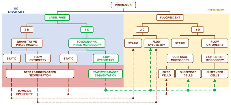
Quantitative Phase Imaging (QPI) has gained popularity in bioimaging because it can avoid the need for cell staining, which in some cases is difficult or impossible. However, as a result, QPI does not provide labelling of various specific intracellular structures. Here we show a novel computational segmentation method based on statistical inference that makes it possible for QPI techniques to identify the cell nucleus. We demonstrate the approach with refractive index tomograms of stain-free cells reconstructed through the tomographic phase microscopy in flow cytometry mode. In particular, by means of numerical simulations and two cancer cell lines, we demonstrate that the nucleus can be accurately distinguished within the stain-free tomograms. We show that our experimental results are consistent with confocal fluorescence microscopy (FM) data and microfluidic cytofluorimeter outputs. This is a significant step towards extracting specific three-dimensional intracellular structures directly from the phase-contrast data in a typical flow cytometry configuration.

摘要

定量相成像(QPI)在生物成像领域已颇受欢迎,因为它无需进行细胞染色,而在某些情况下,细胞染色操作困难甚至无法进行。然而,其结果是,QPI无法对各种特定的细胞内结构进行标记。在此,我们展示了一种基于统计推断的新型计算分割方法,该方法使QPI技术能够识别细胞核成为可能。我们通过流式细胞术模式下的层析相显微镜重建的无染色细胞的折射率断层图来演示该方法。特别是,通过数值模拟和两种癌细胞系,我们证明了在无染色断层图中可以准确区分细胞核。我们表明,我们的实验结果与共聚焦荧光显微镜(FM)数据和微流控细胞荧光计输出结果一致。这是朝着在典型的流式细胞术配置中直接从相衬数据提取特定三维细胞内结构迈出的重要一步。

https://cdn.ncbi.nlm.nih.gov/pmc/blobs/21c6/7613862/18d6c5f51ea0/EMS155286-f005.jpg
https://cdn.ncbi.nlm.nih.gov/pmc/blobs/21c6/7613862/4c39796e1228/EMS155286-f001.jpg
https://cdn.ncbi.nlm.nih.gov/pmc/blobs/21c6/7613862/1d701ad11d91/EMS155286-f002.jpg
https://cdn.ncbi.nlm.nih.gov/pmc/blobs/21c6/7613862/3f060488cdc9/EMS155286-f003.jpg
https://cdn.ncbi.nlm.nih.gov/pmc/blobs/21c6/7613862/666c70fd2d3d/EMS155286-f004.jpg
https://cdn.ncbi.nlm.nih.gov/pmc/blobs/21c6/7613862/18d6c5f51ea0/EMS155286-f005.jpg

相似文献

1
Stain-free identification of cell nuclei using tomographic phase microscopy in flow cytometry.在流式细胞术中使用断层相显微镜对细胞核进行无标记识别。
Nat Photonics. 2022 Dec;16(12):851-859. doi: 10.1038/s41566-022-01096-7. Epub 2022 Nov 10.
2
CellSNAP: a fast, accurate algorithm for 3D cell segmentation in quantitative phase imaging.CellSNAP:一种用于定量相衬成像中三维细胞分割的快速、准确算法。
J Biomed Opt. 2024 Jun;29(Suppl 2):S22706. doi: 10.1117/1.JBO.29.S2.S22706. Epub 2024 Apr 18.
3
CellSNAP: A fast, accurate algorithm for 3D cell segmentation in quantitative phase imaging.CellSNAP:一种用于定量相成像中三维细胞分割的快速、准确算法。
bioRxiv. 2023 Aug 13:2023.07.24.550376. doi: 10.1101/2023.07.24.550376.
4
Acetic acid enabled nuclear contrast enhancement in epi-mode quantitative phase imaging.乙酸可在落射光模式定量相成像中实现细胞核对比度增强。
J Biomed Opt. 2025 Feb;30(2):026501. doi: 10.1117/1.JBO.30.2.026501. Epub 2025 Feb 4.
5
Label-free liquid biopsy through the identification of tumor cells by machine learning-powered tomographic phase imaging flow cytometry.基于机器学习的断层相位成像流式细胞术识别肿瘤细胞的无标记液体活检。
Sci Rep. 2023 Apr 13;13(1):6042. doi: 10.1038/s41598-023-32110-9.
6
Label-Free Intracellular Multi-Specificity in Yeast Cells by Phase-Contrast Tomographic Flow Cytometry.通过相差断层扫描流式细胞术实现酵母细胞中无标记的细胞内多特异性
Small Methods. 2023 Nov;7(11):e2300447. doi: 10.1002/smtd.202300447. Epub 2023 Sep 5.
7
Automatic three-dimensional segmentation of mouse embryonic stem cell nuclei by utilising multiple channels of confocal fluorescence images.利用共聚焦荧光图像的多个通道自动对小鼠胚胎干细胞核进行三维分割。
J Microsc. 2021 Jan;281(1):57-75. doi: 10.1111/jmi.12949. Epub 2020 Aug 8.
8
DryMass: handling and analyzing quantitative phase microscopy images of spherical, cell-sized objects.DryMass:处理和分析球形、细胞大小物体的定量相显微镜图像。
BMC Bioinformatics. 2020 Jun 3;21(1):226. doi: 10.1186/s12859-020-03553-y.
9
Integral refractive index imaging of flowing cell nuclei using quantitative phase microscopy combined with fluorescence microscopy.使用定量相显微镜与荧光显微镜相结合对流动细胞核进行整体折射率成像。
Biomed Opt Express. 2018 Feb 15;9(3):1177-1189. doi: 10.1364/BOE.9.001177. eCollection 2018 Mar 1.
10
Perspective on quantitative phase imaging to improve precision cancer medicine.定量相位成像在提高精准癌症医学中的应用展望。
J Biomed Opt. 2024 Jun;29(Suppl 2):S22705. doi: 10.1117/1.JBO.29.S2.S22705. Epub 2024 Mar 26.

引用本文的文献

1
Drug-Induced Reversible Lysosomal Changes Tracked in Live Cells by Holo-Tomographic Flow Cytometry.通过全断层流式细胞术在活细胞中追踪药物诱导的可逆溶酶体变化
ACS Nano. 2025 Aug 19;19(32):29601-29615. doi: 10.1021/acsnano.5c08530. Epub 2025 Aug 6.
2
From genotype to phenotype: decoding mutations in blasts by holo-tomographic flow cytometry.从基因型到表型:通过全断层流式细胞术解码原始细胞中的突变
Light Sci Appl. 2025 Jul 2;14(1):233. doi: 10.1038/s41377-025-01913-y.
3
Femtosecond laser microfabrication of a fully-integrated optofluidic device for 3D imaging flow cytometry.

本文引用的文献

1
Speeding up reconstruction of 3D tomograms in holographic flow cytometry deep learning.利用深度学习技术加速全息流式细胞术三维断层重建
Lab Chip. 2022 Feb 15;22(4):793-804. doi: 10.1039/d1lc01087e.
2
Label-free multiplexed microtomography of endogenous subcellular dynamics using generalizable deep learning.基于可推广深度学习的无标记多路复用微层析术用于内源性亚细胞动力学研究。
Nat Cell Biol. 2021 Dec;23(12):1329-1337. doi: 10.1038/s41556-021-00802-x. Epub 2021 Dec 7.
3
Dehydration of plant cells shoves nuclei rotation allowing for 3D phase-contrast tomography.
用于三维成像流式细胞术的全集成光流体装置的飞秒激光微加工
Sci Rep. 2025 Apr 8;15(1):11950. doi: 10.1038/s41598-025-93118-x.
4
Neuromorphic-enabled video-activated cell sorting.基于神经形态的视频激活细胞分选
Nat Commun. 2024 Dec 30;15(1):10792. doi: 10.1038/s41467-024-55094-0.
5
Chemistry and properties of fluorescent pyrazole derivatives: an approach to bioimaging applications.荧光吡唑衍生物的化学性质与特性:生物成像应用方法
RSC Adv. 2024 Dec 11;14(53):39230-39241. doi: 10.1039/d4ra07485h. eCollection 2024 Dec 10.
6
High-throughput single-cell sorting by stimulated Raman-activated cell ejection.通过受激拉曼激活细胞喷射进行高通量单细胞分选
Sci Adv. 2024 Dec 13;10(50):eadn6373. doi: 10.1126/sciadv.adn6373. Epub 2024 Dec 11.
7
Three-dimensional isotropic imaging of live suspension cells enabled by droplet microvortices.液滴微涡旋实现活悬浮细胞的三维各向同性成像。
Proc Natl Acad Sci U S A. 2024 Oct 29;121(44):e2408567121. doi: 10.1073/pnas.2408567121. Epub 2024 Oct 22.
8
Long-term three-dimensional high-resolution imaging of live unlabeled small intestinal organoids via low-coherence holotomography.通过低相干全层析成像对活的未标记的小肠类器官进行长期的三维高分辨率成像。
Exp Mol Med. 2024 Oct;56(10):2162-2170. doi: 10.1038/s12276-024-01312-0. Epub 2024 Oct 1.
9
Quantitative phase imaging based on holography: trends and new perspectives.基于全息术的定量相位成像:趋势与新视角。
Light Sci Appl. 2024 Jun 27;13(1):145. doi: 10.1038/s41377-024-01453-x.
10
Dynamic label-free analysis of SARS-CoV-2 infection reveals virus-induced subcellular remodeling.动态无标记分析 SARS-CoV-2 感染揭示了病毒诱导的细胞内重塑。
Nat Commun. 2024 Jun 11;15(1):4996. doi: 10.1038/s41467-024-49260-7.
植物细胞脱水促使细胞核旋转,从而实现三维相衬断层扫描。
Light Sci Appl. 2021 Sep 15;10(1):187. doi: 10.1038/s41377-021-00626-2.
4
Label-free colorectal cancer screening using deep learning and spatial light interference microscopy (SLIM).使用深度学习和空间光干涉显微镜(SLIM)进行无标记结直肠癌筛查。
APL Photonics. 2020 Apr;5(4). doi: 10.1063/5.0004723. Epub 2020 Apr 28.
5
Three-Dimensional Quantitative Intracellular Visualization of Graphene Oxide Nanoparticles by Tomographic Flow Cytometry.通过断层流式细胞术对石墨烯氧化物纳米粒子进行三维定量细胞内可视化。
Nano Lett. 2021 Jul 28;21(14):5958-5966. doi: 10.1021/acs.nanolett.1c00868. Epub 2021 Jul 7.
6
Holotomography: Refractive Index as an Intrinsic Imaging Contrast for 3-D Label-Free Live Cell Imaging.体全息术:折射率作为一种固有成像对比用于三维无标记活细胞成像。
Adv Exp Med Biol. 2021;1310:211-238. doi: 10.1007/978-981-33-6064-8_10.
7
Rolling angle recovery of flowing cells in holographic tomography exploiting the phase similarity.利用相位相似性实现全息层析术中流动细胞的滚动角恢复。
Appl Opt. 2021 Feb 1;60(4):A277-A284. doi: 10.1364/AO.404376.
8
Phase imaging with computational specificity (PICS) for measuring dry mass changes in sub-cellular compartments.相位成像与计算特异性(PICS)用于测量亚细胞区室中干物质变化。
Nat Commun. 2020 Dec 7;11(1):6256. doi: 10.1038/s41467-020-20062-x.
9
Machine learning of diffraction image patterns for accurate classification of cells modeled with different nuclear sizes.基于不同核大小模型的细胞衍射图像模式的机器学习实现精确分类。
J Biophotonics. 2020 Sep;13(9):e202000036. doi: 10.1002/jbio.202000036. Epub 2020 Jul 3.
10
High-resolution 4-D acquisition of freely swimming human sperm cells without staining.高分辨率 4D 采集自由游动的人类精子细胞,无需染色。
Sci Adv. 2020 Apr 10;6(15):eaay7619. doi: 10.1126/sciadv.aay7619. eCollection 2020 Apr.