Jiang Xin'ang, Hu Jun, Jiang Qinling, Zhou Taohu, Yao Fei, Sun Yi, Liu Qingyang, Zhou Chao, Shi Kang, Lin Xiaoqing, Li Jie, Li Yueze, Jin Qianxi, Tu Wenting, Zhou Xiuxiu, Wang Yun, Xin Xiaoyan, Liu Shiyuan, Fan Li
Department of Radiology, Second Affiliated Hospital of Naval Medical University, Shanghai, China.
Department of Radiology, Nanjing Drum Tower Hospital, The Affiliated Hospital of Nanjing University Medical School, Nanjing, China.
J Thorac Dis. 2024 Sep 30;16(9):5591-5603. doi: 10.21037/jtd-24-544. Epub 2024 Sep 6.
Coronavirus disease 2019 (COVID-19) still poses a threat to people's physical and mental health. We proposed a new semi-quantitative visual classification method for COVID-19, and this study aimed to evaluate the clinical usefulness and feasibility of lung field-based severity score (LFSS).
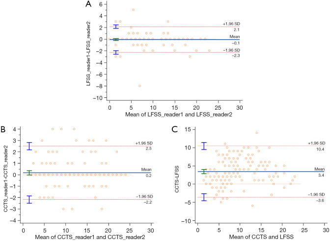
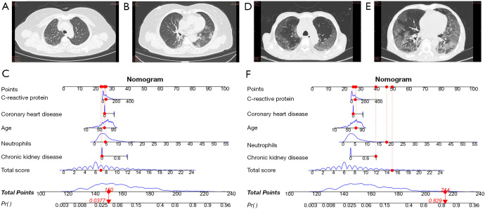
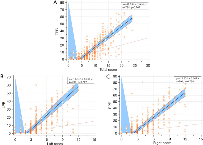
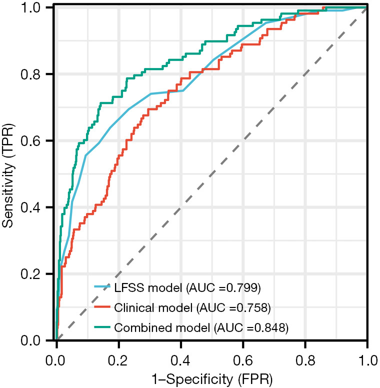
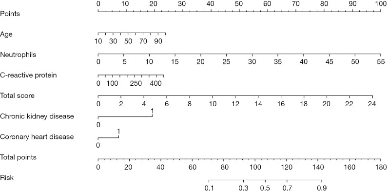
This retrospective study included 794 COVID-19 patients from two hospitals in China between December 2022 and January 2023. Six lung fields on the axial computed tomography (CT) were defined. LFSS and eighteen clinical characteristics were evaluated. LFSS was based on summing up the parenchymal opacification involving each lung field, which was scored as 0 (0%), 1 (1-24%), 2 (25-49%), 3 (50-74%), or 4 (75-100%), respectively (range of LFSS from 0 to 24). Total pneumonia burden (TPB) was calculated using the U-net model. The correlation between LFSS and TPB was analyzed. After performing logistic regression analysis, an LFSS-based model, clinical-based model and combined model were developed. Receiver operating characteristic curves were used to evaluate and compare the performance of three models.
LFSS, age, chronic liver disease, chronic kidney disease, white blood cell, neutrophils, lymphocytes and C-reactive protein differed significantly between the non-critical and critical group (all P<0.05). There was a strong positive correlation of LFSS and TPB (Pearson correlation coefficient =0.767, P<0.001). The area under curves of LFSS-based model, clinical-based model and combined model were 0.799 [95% confidence interval (CI): 0.770-0.827], 0.758 (95% CI: 0.727-0.788), and 0.848 (95% CI: 0.821-0.872), respectively.
The LFSS derived from chest CT may be a potential new tool to help identify COVID-19 patients at high risk of progressing to critical disease.
2019冠状病毒病(COVID-19)仍然对人们的身心健康构成威胁。我们提出了一种新的用于COVID-19的半定量视觉分类方法,本研究旨在评估基于肺野的严重程度评分(LFSS)的临床实用性和可行性。
这项回顾性研究纳入了2022年12月至2023年1月期间来自中国两家医院的794例COVID-19患者。定义了轴向计算机断层扫描(CT)上的六个肺野。评估了LFSS和18项临床特征。LFSS基于对每个肺野实质混浊情况进行求和,分别评分为0(0%)、1(1 - 24%)、2(25 - 49%)、3(50 - 74%)或4(75 - 100%)(LFSS范围为0至24)。使用U-net模型计算总肺炎负担(TPB)。分析了LFSS与TPB之间的相关性。进行逻辑回归分析后,建立了基于LFSS的模型、基于临床的模型和联合模型。使用受试者操作特征曲线评估和比较三种模型的性能。
非重症组和重症组之间的LFSS、年龄、慢性肝病、慢性肾病、白细胞、中性粒细胞、淋巴细胞和C反应蛋白存在显著差异(均P<0.05)。LFSS与TPB呈强正相关(Pearson相关系数 = 0.767,P<0.001)。基于LFSS的模型、基于临床的模型和联合模型的曲线下面积分别为0.799 [95%置信区间(CI):0.770 - 0.827]、0.758(95% CI:0.727 - 0.788)和0.848(95% CI:0.821 - 0.872)。
源自胸部CT的LFSS可能是一种潜在的新工具,有助于识别有进展为重症疾病高风险的COVID-19患者。