Kim Bo Kyoung, Goncharov Tatiana, Archaimbault Sébastien A, Roudnicky Filip, Webster Joshua D, Westenskow Peter D, Vucic Domagoj
Department of Ophthalmology Discovery, Pharmaceutical Research and Early Development, Roche Innovation Center Basel, F. Hoffmann-La Roche Ltd, Basel, Switzerland.
Institute of Chemical Sciences and Engineering (ISIC), École Polytechnique Fédérale de Lausanne (EPFL), Lausanne, Switzerland.
Cell Death Differ. 2025 Feb;32(2):353-368. doi: 10.1038/s41418-024-01390-7.
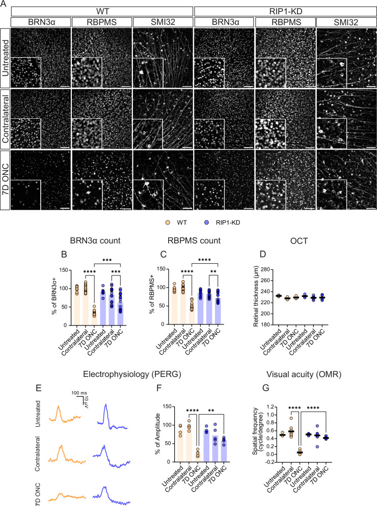
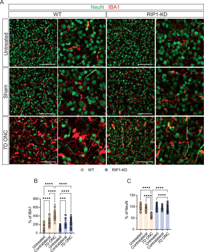
Receptor-interacting protein 1 (RIP1, RIPK1) is a critical mediator of multiple signaling pathways that promote inflammatory responses and cell death. The kinase activity of RIP1 contributes to the pathogenesis of a number of inflammatory and neurodegenerative diseases. However, the role of RIP1 in retinopathies remains unclear. This study demonstrates that RIP1 inhibition protects retinal ganglion cells (RGCs) in preclinical glaucoma models. Genetic inactivation of RIP1 improves RGC survival and preserves retinal function in the preclinical glaucoma models of optic nerve crush (ONC) and ischemia-reperfusion injury (IRI). In addition, the involvement of necroptosis in ONC and IRI glaucoma models was examined by utilizing RIP1 kinase-dead (RIP1-KD), RIP3 knockout (RIP3-KO), and MLKL knockout (MLKL-KO) mice. The number of RGCs, retinal thickness, and visual acuity were rescued in RIP1-kinase-dead (RIP1-KD) mice in both models, while wild-type (WT) mice experienced significant retinal thinning, RGC loss, and vision impairment. RIP3-KO and MLKL-KO mice showed moderate protective effects in the IRI model and limited in the ONC model. Furthermore, we confirmed that a glaucoma causative mutation in optineurin, OPTN-E50K, sensitizes cells to RIP1-mediated inflammatory cell death. RIP1 inhibition reduces RGC death and axonal degeneration following IRI in mice expressing OPTN-WT and OPTN-E50K variant mice. We demonstrate that RIP1 inactivation suppressed microglial infiltration in the RGC layer following glaucomatous damage. Finally, this study highlights that human glaucomatous retinas exhibit elevated levels of TNF and RIP3 mRNA and microglia infiltration, thus demonstrating the role of neuroinflammation in glaucoma pathogenesis. Altogether, these data indicate that RIP1 plays an important role in modulating neuroinflammation and that inhibiting RIP1 activity may provide a neuroprotective therapy for glaucoma.
受体相互作用蛋白1(RIP1,RIPK1)是多种促进炎症反应和细胞死亡的信号通路的关键介质。RIP1的激酶活性参与多种炎症性和神经退行性疾病的发病机制。然而,RIP1在视网膜病变中的作用仍不清楚。本研究表明,在临床前青光眼模型中,抑制RIP1可保护视网膜神经节细胞(RGCs)。在视神经挤压(ONC)和缺血再灌注损伤(IRI)的临床前青光眼模型中,RIP1的基因失活可改善RGC的存活并保留视网膜功能。此外,通过使用RIP1激酶失活(RIP1-KD)、RIP3基因敲除(RIP3-KO)和混合谱系激酶结构域样蛋白(MLKL)基因敲除(MLKL-KO)小鼠,研究了坏死性凋亡在ONC和IRI青光眼模型中的作用。在两种模型中,RIP1激酶失活(RIP1-KD)小鼠的RGC数量、视网膜厚度和视力均得到挽救,而野生型(WT)小鼠则出现明显的视网膜变薄、RGC丢失和视力损害。RIP3-KO和MLKL-KO小鼠在IRI模型中显示出中等程度的保护作用,而在ONC模型中作用有限。此外,我们证实,青光眼致病基因视紫质神经元(optineurin)中的突变OPTN-E50K使细胞对RIP1介导的炎症性细胞死亡敏感。在表达OPTN-WT和OPTN-E50K变异体的小鼠中,抑制RIP1可减少IRI后RGC的死亡和轴突退变。我们证明,RIP1失活可抑制青光眼性损伤后RGC层中的小胶质细胞浸润。最后,本研究强调,人类青光眼视网膜中肿瘤坏死因子(TNF)和RIP3的mRNA水平升高,且有小胶质细胞浸润,从而证明了神经炎症在青光眼发病机制中的作用。总之,这些数据表明,RIP1在调节神经炎症中起重要作用,抑制RIP1活性可能为青光眼提供一种神经保护疗法。